
The relief of symptoms remains the primary force driving antireflux surgery in patients with Barrett's esophagus. Healing of esophageal mucosal injury and the prevention of disease progression are important secondary goals. In this regard, patients with Barrett's esophagus are no different than the broader population of patients with GE reflux. Antireflux surgery should be considered when patient factors suggest severe disease or predict the need for long-term medical management, both of which are almost always true in patients with Barrett's esophagus.
PPI therapy, both to relieve symptoms and to control any coexistent esophagitis or stricture, is an acceptable treatment option in patients with Barrett's esophagus. Once initiated, however, most patients with Barrett's will require lifelong treatment. Complete control of reflux with PPI therapy can be difficult, however, as has been highlighted by studies of acid breakthrough while on therapy. Ablation trials have shown that mean doses of 56 mg of omeprazole are necessary to normalize 24-hour esophageal pH studies. Antireflux surgery likely results in more reproducible and reliable elimination of reflux of both acid and duodenal content, although long-term outcome studies suggest that as many as 25% of patients postfundoplication have persistent pathologic esophageal acid exposure confirmed by 24-hour pH studies.58
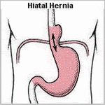
An important consideration is that patients with Barrett's esophagus generally have severe GERD, with its attendant sequelae such as large hiatal hernia, stricture, shortened esophagus, and poor motility. These anatomic and physiologic features make successful antireflux surgery a particular challenge in this population. Indeed, recent data suggest that antireflux surgery in patients with Barrett's esophagus may not be as successful in the long term as in those without Barrett's.
Studies focusing on the symptomatic outcome following antireflux surgery in patients with Barrett's esophagus document excellent to good results in 72% to 95% of patients at 5 years following surgery The outcome of laparoscopic Nissen fundoplication in patients with Barrett's esophagus has been assessed at 1 to 3 years after surgery.Reflux symptoms were absent postoperatively in 79% of the patients. Postoperative 24-hour pH was normal in 17 of 21 (81%) patients. Ninety-nine percent of the patients considered themselves cured or improved, and 97% were satisfied with the surgery.
Friday, May 23, 2008
 Treatment of Barrett's esophagus
Tuesday, May 20, 2008
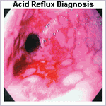
What is Barrett's esophagus?
Barrett's esophagus is the metaplastic complication of Acid Reflux Disease.
It is the condition whereby the tubular esophagus is lined with columnar epithelium rather than squamous epithelium was first described by Norman Barrett in 1950. He incorrectly believed it to be congenital in origin. It is now realized that it is an acquired abnormality, occurring in 7% to 10% of patients with GERD, and represents the end stage of the natural history of this disease. It is also understood to be distinctly different from the congenital condition in which islands of mature gastric columnar epithelium are found in the upper half of the esophagus.
The definition of Barrett's esophagus has evolved considerably over the past decade. Traditionally, Barrett's esophagus was identified by the presence of any columnar mucosa extending at least 3 cm into the esophagus. Recent data indicating that specialized intestinal-type epithelium is the only
Recent studies suggest that the metaplastic process at the GE junction may begin by conversion of distal esophageal squamous mucosa to cardiac-type epithelium, heretofore presumed to be a normal finding. This is likely due to exposure of the distal esophagus to excess acid and gastric contents via prolapse of esophageal squamous mucosa into the gastric environment. This results in inflammatory changes at the GE junction or a metaplastic process, both of which may result in the loss of muscle function and a mechanically defective sphincter allowing free reflux with progressively higher degrees of mucosal injury. Intestinal metaplasia within the sphincter may result, as in Barrett's metaplasia of the esophageal body. This mechanism is supported by the finding that as the severity of GERD progresses, the length of columnar lining above the anatomic GE junction is increased.
Friday, May 16, 2008
Acid Reflux Disease Complications
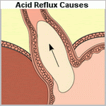
The complications of GE reflux result from the damage inflicted by gastric juice on the esophageal mucosa. Mucosal complications, include esophagitis and stricture. The prevalence and severity of complications is related to the degree of loss of the GE barrier, defects in esophageal clearance, and the content of refluxed gastric juice.
The potential injurious components that reflux into the esophagus include gastric secretions, such as acid and pepsin, biliary and pancreatic secretions that regurgitate from the duodenum into the stomach, and toxic compounds generated in the mouth, esophagus, and stomach by the action of bacteria on dietary substances.
Studies have shown that acid alone does minimal damage to the esophageal mucosa, but the combination of acid and pepsin is highly deleterious. Hydrogen ion injury to the esophageal squamous mucosa occurs only at a pH below 2. In acid refluxate, the enzyme pepsin appears to be the major injurious agent. Similarly, the reflux of duodenal juice alone does little damage to the mucosa, whereas the combination of duodenal juice and gastric acid is particularly noxious. Reflux of bile and pancreatic enzymes into the stomach can either protect against or augment esophageal mucosal injury. For instance, the reflux of duodenal contents into the stomach may prevent the development of peptic esophagitis in a patient whose gastric acid secretion maintains an acid environment, because the bile salts would attenuate the injurious effect of pepsin and the acid would inactivate the trypsin. Such a patient would have bile-containing acid gastric juice that, when refluxed, would irritate
the esophageal mucosa but cause less esophagitis than if it were acid gastric juice containing pepsin. In contrast, the reflux of duodenal contents into the stomach of a patient with limited gastric acid secretion can result in esophagitis, because the alkaline intragastric environment would support optimal trypsin activity, and the soluble bile salts with a high pKa would potentiate the enzyme's effect. Hence, duodenal-gastric reflux and the acid secretory capacity of the stomach interrelate by altering the pH and enzymatic activity of the refluxed gastric juice to modulate the injurious effects of enzymes on the esophageal mucosa.
This disparity in injury caused by acid and bile alone, as opposed to the gross esophagitis caused by pepsin and trypsin, provides an explanation for the poor correlation between the symptom of heartburn and endoscopic esophagitis. The reflux of acid gastric juice contaminated with duodenal contents could break the esophageal mucosal barrier, irritate nerve endings in the papillae close to the luminal surface, and cause severe heartburn. Despite the presence of intense heartburn, the bile salts present would inhibit pepsin, the acid pH would inactivate trypsin, and the patient would have little or no gross evidence of esophagitis. In contrast, the patient who refluxed alkaline gastric juice may have minimal heartburn because of the absence of hydrogen ions in the refluxate but have endoscopic esophagitis because of the bile salt potentiation of trypsin activity on the esophageal mucosa. This is supported by recent clinical studies which indicate that the presence of alkaline reflux is associated with the development of mucosal injury.
Although numerous studies have suggested the reflux of duodenal contents into the esophagus in patients with GERD, few have measured this directly. The components of duodenal juice thought to be most damaging are the bile acids and, as such, they have been the most commonly studied. Most studies shown that, patients with GERD have greater and more concentrated bile acid exposure to the esophageal mucosa than do normal subjects. This increased exposure occurs most commonly during the supine period while asleep and during the upright period following meals. Most studies have identified the glycine conjugates of cholic, deoxycholic, and chenodeoxycholic acids as the predominant bile acids aspirated from the esophagus of patients with GERD, although appreciable amounts of taurine conjugates of these bile acids were also found. Other bile salts were identified but in small concentrations. This is as one would expect because glycine conjugates are three times more prevalent than taurine conjugates in normal human bile.
The potentially injurious action of toxic compounds either ingested or newly formed on the mucosa of the GE junction and distal esophagus has long been postulated. Investigators have recently shown that dietary nitrate consumed in the form of green vegetables and food contaminated by nitrate-containing fertilizers results in the generation of nitric oxide at the GE junction in concentrations high enough to be potentially mutagenic. Previous studies have shown that nitrate ingested in food is reabsorbed in the small bowel with approximately 25% resecreted into the mouth via the salivary glands. Oral bacteria chemically transform the relatively innocuous nitrate to the more toxic nitrite, which is swallowed and subsequently converted to nitric oxide and other toxic nitroso-compounds by acid and ascorbic acid in the stomach. Whether this mechanism in fact contributes to injury and or neoplastic transformation in the upper stomach, GE junction, and distal esophagus is currently unknown.
Tuesday, May 13, 2008
Anti reflux Surgery Complications
Postoperative complications were found to occur in approximately 8% of patients, with the rate of conversion to an open procedure of about 4%. The most common perioperative complication was early wrap herniation (1.3%), defined as occurring within 48 hours of surgery. This is one complication that may be more common with the laparoscopic than open approach. The explanation for this is unclear but may be related to the opening of tissue planes by the pneumoperitoneum and the reduced tendency for adhesion formation after laparoscopic compared to open surgery. In an attempt to eliminate this complication, most surgeons routinely perform a crural repair.
Both pneumothorax and pneumomediastinum have been reported. The occurrence of pneumothorax is related to breach of either pleural membrane, usually the left, during the hiatal dissection. Chest drain insertion is usually not required because accumulated carbon dioxide rapidly dissipates following release of pneumoperitoneum by a combination of positive pressure ventilation and absorption.
As with any laparoscopic procedure, instrumental perforation of the hollow viscera may occur. Early esophageal perforation may arise during passage of the bougie, during the retroesophageal dissection, or during suture pull-through. Late esophageal perforation is related to diathermy injury at the time of mobilization. Gastric perforations usually resulted from excessive traction on the fundus for retraction purposes. Recognition of the problem at the time of surgery requires repair, which may be performed either laparoscopically or by an open technique.
Hemorrhage during the course of laparoscopic fundoplication usually arises from the short gastric vessels or spleen. Rarer causes include retractor trauma to the liver, injury to the left inferior phrenic vein, an aberrant left hepatic vein, or the inferior vena cava. Cardiac tamponade as a result of right ventricular trauma has also been reported. Major vascular injury mandates immediate conversion to an open procedure to achieve hemostasis.
Acid reflux disease facts
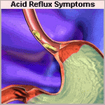
The diagnosis of GE reflux based on symptoms alone is correct in only approximately two thirds of patients because the symptoms are often nonspecific and can be caused by other conditions.
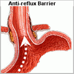
The three characteristics of the LES that maintains its resistance or barrier function to intragastric and intraabdominal pressure challenges are pressure, overall length, and length exposed to the positive pressure environment of the abdomen.
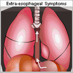
An important complication of GE reflux is the development of reflux-induced respiratory symptoms either with or without heartburn.
Antireflux surgery improves respiratory symptoms in nearly 90% of children and 70% of adults with asthma and reflux disease.
Factors predisposing to the development of Barrett's esophagus include early-onset GERD, abnormal lower esophageal and esophageal body physiology, and mixed reflux of gastric and duodenal contents into the esophagus.
PPI therapy, both to relieve symptoms and to control esophagitis, is an acceptable treatment, although most patients will require life-long treatment.
Progression of nondysplastic Barrett's epithelium occurs with 5% to 10% of patients per year progressing to dysplasia and 0.5% to 1% per year progressing to cancer
The standard of care of the treatment of confirmed Barrett's esophagus with high-grade dysplasia is esophagectomy because approximately 50% will harbor invasive cancer
Three factors predictive of a successful outcome following antireflux surgery are (a) an abnormal score on 24-hour esophageal pH monitoring; (b) the presence of typical symptoms of GERD, namely heartburn or regurgitation; and (c) symptomatic improvement in response to acid-suppression therapy prior to surgery.
Saturday, May 3, 2008
Endoscopic investigation of gastro-esophageal reflux
The development of fibre-optic endoscopy revolutionized the ability to investigate the gastrointestinal tract. Flexible endoscopes often have a diameter of less than 1 cm, with a control head and a flexible shaft with a manoeuvrable tip. The head is connected to a light source and can transmit images to a video image screen.
All endoscopes have multiple small lumens allowing transmission of air and water and for suction. The suction channel can also be used for the passage of interventional devices, for example biopsy forceps. The ability to transmit air (insufflation) allows the endoscopist to inflate the lumen to obtain optimal views. The water channel provides a means of washing mucosal surfaces, and suction maybe used to remove pools of fluid within the gastrointestinal tract, thus ensuring that all mucosal surfaces are inspected.
Diagnostic indications include the investigation of gastro-esophageal reflux (diagnostic investigation or surveillance endoscopy of Barrett's esophagus).
Therapeutic procedures that can be undertaken include mucosal biopsy, stent insertion for strictures, and dilatation of strictures.
Patient preparation
Informed consent is required, and patients are fasted for 4-6 hours prior to the procedure. Although most patients do not require sedation, the choice is offered and discussed. Sedation involves the use of a short-acting benzodiazepine which provides a sedative and amnesic effect. Monitoringis required (pulse oximetry) with sedation due to the risk of respiratory depression. Antibiotic prophylaxis is administered to patients with heart valve disease to prevent bacterial endocarditis.
Procedure
A mouthguard is used. The endoscope is introduced into the pharynx, then the oesophagus. Patients may retch during this procedure. The endoscope is progressively introduced to inspect the oesophagus, stomach and proximal small bowel (duodenum).
Complications
The overall complication rate is approximately 1 per 1000 with a mortality rate of approximately 1 per 25000. Complications include bleeding, perforation and respiratory arrest (a complication of sedation).
Post procedure care
Patients are monitored in a recovery area until safe for discharge.

