
Once GERD is suspected or thought to be responsible for asthma symptoms, treatment may be with either prolonged PPI (proton pump inhibitor) therapy or anti reflux surgery. A 3- to 6-month trial of high-dose PPI therapy [twice a day (b.i.d.) or three times a day (t.i.d.) dosing] may help confirm (by virtue of symptom resolution) that GERD is partly or completely responsible for the asthma symptoms. The persistence of symptoms despite PPI treatment, however, does not necessarily rule out GERD as a potential contributor.
Based on reported observations, relief of asthma symptoms can be anticipated for 25% to 50% of patients with GERD asthma treated with anti reflux medications.
Fewer than 15%, however, can be expected to have objective improvements in their pulmonary function. The reason for this apparent paradox may be that most studies employed relatively short courses of anti reflux therapy (less than 3 months). This time period may have been sufficient for symptomatic improvement but insufficient for recovery of pulmonary function. The chances of success with medical treatment are likely directly related to the extent of GERD elimination. The conflicting findings of reports of anti reflux therapy may well be to the result of inadequate control of GERD in some studies. The literature indicates that anti reflux surgery improves asthma symptoms in nearly 90% of children and 70% of adults with asthma and GERD. Improvements in pulmonary function were demonstrated in around one third of patients. Comparison of the results of uncontrolled studies of each form of therapy and the evidence from the two randomized controlled trials of medical versus surgical therapy indicate that fundoplication is the most effective therapy for GERD asthma. The superiority of the surgical anti reflux barrier over medical therapy is probably most noticeable in the supine posture, which corresponds with the period of acid breakthrough with PPI therapy and is the time in the circadian cycle when asthma symptoms and peak expiratory flow rates are at their worst.
It is also important to realize that, in asthmatic patients with a non reflux induced motility abnormality of the esophageal body, performing an anti reflux operation may not prevent the aspiration of orally regurgitated, swallowed liquid or food. This can result in asthma symptoms and airway irritation that may elicit an asthmatic reaction. This factor may explain why surgical results appear to be better in children than adults, because disturbance of esophageal body motility is more likely in adult patients.
Thursday, March 20, 2008
 GERD Asthma Treatment
Monday, March 17, 2008
GERD Asthma
Two mechanisms have been proposed as the pathogenesis of GERD asthma. The first, the so-called reflux theory, maintains that asthma is the result of the aspiration of gastric contents. The second or reflex theory maintains that vagally mediated bronchoconstriction follows acidification of the lower esophagus. The evidence supporting a reflux mechanism is fivefold. First, clinical studies have documented a strong correlation between idiopathic pulmonary fibrosis and hiatal hernia. The presence of GERD was shown to be highly associated with several pulmonary diseases, not only asthma, in recent studies. Second, pathologic acid exposure in the proximal esophagus is often identified in patients with asthma and reflux disease. Third, scintigraphic studies have shown aspiration of ingested radioisotope in some patients with reflux and respiratory symptoms. Fourth, simultaneous tracheal and esophageal pH monitoring in patients with reflux disease has documented tracheal acidification in concert with esophageal acidification. Finally, animal studies have shown that tracheal instillation of hydrochloric acid profoundly increases airways resistance.
A reflex mechanism is primarily supported by the fact that bronchoconstriction occurs following the infusion of acid into the lower esophagus. This can be explained by the common embryologic origin of the tracheoesophageal tract and its shared vagal innervation. Second, patients with asthma and pathologic distal esophageal acid exposure but normal proximal esophageal acid exposure may experience an improvement in their asthma after antireflux therapy.
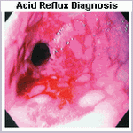
The primary challenge in implementing treatment for reflux-associated asthma lies in establishing the diagnosis. In patients with predominantly typical reflux symptoms and secondary respiratory complaints, the diagnosis may be straightforward. However, in a substantial number of patients with GERD asthma, the respiratory symptoms dominate the clinical scenario. GE reflux in these patients is often silent and is uncovered only when investigation is initiated. A high index of suspicion is required, notably in patients with poorly controlled asthma despite appropriate bronchodilator therapy. Supportive evidence for the diagnosis can be gleaned from endoscopy and stationary esophageal manometry. Endoscopy may show erosive esophagitis or Barrett's esophagus. Manometry may indicate a hypotensive LES or ineffective body motility, defined by 30% or more contractions in the distal esophagus of less than 30 mm Hg in amplitude.
The gold standard for the diagnosis of GERD asthma is ambulatory dual-probe pH monitoring. One probe is positioned in the distal esophagus and the other at a more proximal location. Sites for proximal probe placement have included the trachea, pharynx, and proximal esophagus. Most authorities would agree that the proximal esophagus is the preferred site for proximal probe placement. Although ambulatory esophageal pH monitoring allows a direct correlation between esophageal acidification and respiratory symptoms, the chronologic relationship between reflux events and bronchoconstriction is complex.
Friday, March 14, 2008
What is (GERD)?
(GERD) Gastro-esophageal reflux disease
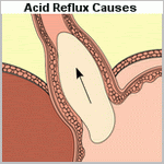
Spontaneous gastro-esophageal reflux may occur as a normal event, but gastro-esophageal reflux disease (GERD) is defined as symptoms (heartburn) and/or tissue damage caused by retrograde flow of gastric contents into the esophagus.
Epidemiology
Symptoms of GERD have been reported in approximately 44% of a surveyed population, but the precise prevalence varies according to the definition of the disease. The incidence increases with age and peaks at the age of 55-64 years, with an approximately equal gender distribution.
Pathology
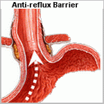
The normal anti-reflux mechanism involves both anatomical and physiological factors: the lower esophageal sphincter, angle of His, crura of the diaphragm, mucosal rosette, swallowed saliva (lubrication and neutralization of acid), antegrade esophageal peristalsis and normal gastric motility/emptying.
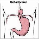
A mild degree of reflux is experienced in most normal individuals, but a major cause of significant GERD is inappropriate transient lower esophageal sphincter relaxations (not preceded by a primary propagated esophageal contraction initiated by swallowing). Other causes include hiatus hernia and delayed gastric emptying.
Although a greater proportion of patients with symptoms of GERD smoke and consume alcohol, clinical studies confirm that traditional risk factors of increasing age, male sex, smoking and alcohol consumption are not strongly associated with symptoms of GERD.
Scope of disease
The majority have mild disease without esophagitis (non-erosive reflux disease). Symptoms can arise with minimal gastric reflux due to increased sensitivity of the esophagus to acid. Moderate disease is associated with esophagitis, and severe chronic disease results in the development of Barrett's esophagus (defined as metaplastic columnar degeneration above the gastro-esophageal junction). This is a premalignant condition, with a 0.5% per patient-year risk of malignant change to adenocarcinoma.
Severe or prolonged reflux can lead to ulceration, bleeding or perforation. Healing occurs by fibrosis and may lead to stricture formation. Severe reflux may also produce a hoarse voice from laryngitis. If the refluxate is aspirated, pneumonia may develop.
Clinical features
Most patients complain of heartburn, characterized by retrosternal burning pain, precipitated by meals. It may be aggravated by posture (lying flat, stooping, bending forwards) and conditions that raise intra-abdominal pressure (sneezing and coughing). It is often relieved by antacids and may be associated with water brash, the excessive secretion of saliva preceding reflux.
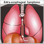
Atypical symptoms include back pain, cardiac-type chest pain, chronic wheeze, nocturnal 'asthma' or recurrent chest infections (due to aspiration), sore throat, hoarse voice, halitosis or dental decay. Dysphagia may occur with esophageal strictures from chronic disease.
Initial investigations
In general, the history can lead to a confident diagnosis of GERD and initial treatment can commence for uncomplicated cases. Older patients, or those who experience weight loss, dysphagia or hematemesis will require further investigations.
Further investigations
Upper gastrointestinal endoscopy
esophagoscopy is appropriate for patients with symptoms suggestive of complications (weight loss, dysphagia, hematemesis), when the diagnosis is unclear, when symptoms cannot be adequately controlled on medical therapy, or for screening for Barrett's epithelium (in patients over50 with chronic symptoms).
esophageal abnormalities that can be diagnosed at endoscopy include hiatus hernia, esophagitis, Barrett's esophagus, strictures and tumours. The severity of reflux esophagitis can also be assessed at endoscopy, and biopsies can be taken. GERD cannot be excluded by a normal endoscopy.
24-hour pH measurement
Intra-esophageal pH measurement may be required to diagnose GERD if the initial endoscopy is normal or if symptoms do not respond to medical therapy. It is usually performed as an ambulatory test over a 24-hour period, facilitating the diagnosis of GERD by assessment of the duration, frequency and severity of reflux attacks in relation to the pH of the esophagus. It does not provide any information as to the etiology of GERD and may be normal (false negative) in patients with significant bile or volume reflux.
esophageal manometry
Esophageal manometry is a useful investigation to assess esophageal function when planning surgical intervention, especially if achalasia and other esophageal motility disorders are suspected. It is possible to determine the length and position of the lower esophageal sphincter and to measure the sphincter pressure. A short or weak lower esophageal sphincter is often a major contributing cause of reflux esophagitis.
Initial management
Lifestyle modification
Suggestions that may improve symptoms include weight loss for obese patients, stopping smoking, frequent small meals, avoiding foods that are known to precipitate symptoms, avoiding eating for several hours before bedtime, and raising the head of the bed. However, evidence for improvement associated with these measuresis scarce.
Medical management
There are three pharmacological approaches to controlling symptoms of GERD: neutralization of acid (antacids and alginates), reduction in acid production (H2-receptor blockers, proton pump inhibitors) and increasing gastrointestinal motility (metoclopramide).
Step-up therapy
Most agents for initial therapy are available without prescription, and pharmacists offer the first-line management of GERD in a step-up regimen (starting from the simplest, most cost-effective agent).
Antacids and alginates
Antacids, such as bicarbonate, act by reducing the acidity within the stomach and lower esophagus. Alginate preparations (such as Gaviscon) act by coating the top of the gastric contents to reduce the effect of acid on the esophageal mucosa. Both these agents are effective, but the effects are often short-lived and suitable only for patients with mild symptoms.
H2-receptor antagonists
H2-receptor antagonists (e.g. cimetidine, ranitidine) block the H2-receptors in gastric mucosa, leading to a marked reduction in gastric acid production. Initial symptomatic relief is experienced in approximately 60%.
Step-down therapy
Patients who seek medical attention may already be on a combination of antacids, alginates and H2-receptor antagonists. Current consensus favours step-down therapy (starting with the most effective agent), as it is associated with greater symptomatic relief and fewer treatment failures and physician consultations.
Proton pump inhibitors
The proton pump inhibitors (e.g. omeprazole, lansoprazole) act by blocking the proton pump within the gastric mucosa, almost completely abolishing gastric acid production. Initial symptomatic relief is experienced in approximately 83%.6 An initial 2-4-week course is recommended followed by maintenance therapy. Failure to respond to initial therapy is an indication for further investigations (endoscopy, pH studies) to confirm and assess the severity of the disease.
Prokinetic agents
Due to the superiority of proton pump inhibitors, the role of prokinetic agents (metoclopramide) is ill defined. Prokinetic agents are usually reserved for use in combination with proton pump inhibitor for step-up therapy, or in combination with an H2-receptor blocker for long-term maintenance step-down therapy.
Maintenance therapy
After 6 months of initial therapy 75% of patients experience symptom relapse. A trial of withdrawal of drug therapy may be initiated but the majority will require maintenance therapy either by step-down treatment (long-term H2-receptor blocker) or intermittent on-demand proton pump inhibitor therapy, a 2-4-week course each time symptoms recur.
Surgical management
Before considering anti-reflux surgery, upper gastrointestinal endoscopy, 24-hour pH studies and esophageal manometry are required for confirmatory diagnosis and documentation of the presence and severity of reflux.
Anti-reflux surgery provides effective long-term treatment of GERD with symptomatic control equivalent to medical therapy but lower rates of esophagitis.10 The indications for surgery are failure of medical treatmentor development of complications (ulceration, strictures, Barrett's esophagus or respiratory complications). Relative indications include volume reflux (i.e. excessive volume rather than acid content) and patient preference.
Anti-reflux surgery has a 90% initial success rate. Complications of surgery include dysphagia (usually short-lived)in about 10%, inability to belch, the so-called 'gas bloat syndrome' (20%) and excessive flatus.
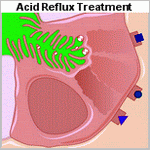
Nissen fundoplication
The most common operation is the Nissen (360 degree) fundoplication, which can be performed via an abdominal incision (midline laparotomy) or laparoscopically. The hiatus of the diaphragm is repaired, the lower esophagus is mobilized, then the greater curvature of the stomach via division of the short gastric arteries. The fundus of the stomach is wrapped completely around the lower esophagus.
To reduce the risk of dysphagia, various partial fundoplications have been described, including the anterior partial (Watson), posterior partial (Toupet) and Lind (270 degree) subtotal fundoplications.
Collis-Nissen procedure
This uncommon procedure is reserved for situations where the gastro-esophageal junction cannot be reduced below the diaphragm. An abdominal approach is used and a linear stapler is used to 'lengthen' the esophagus by incorporating the cardia of the stomach prior to creating the wrap.
Management of benign strictures
A barium swallow is often performed to delineate the site and extent of the stricture, followed by endoscopy and biopsies to determine the etiology. The treatment of benign strictures consists of bougie dilatation, and several sessions may be required for resistant strictures. Further treatment options include resection or bypass of the stricture and long-term intubation of the esophagus.
Following esophageal dilatation, the patient is keptnil by mouth if symptoms such as chest or back pain or surgical emphysema develop. A chest film is performed to exclude a pneumomediastinum, which will suggest esophageal perforation.
Prognosis
GERD is a chronic relapsing condition. Long-term studies report 10-year recurrence rates (based on use of anti-reflux medications) in 62% of surgically treated and 92% of medically treated patients. If Barrett's esophagus develops, regular endoscopic surveillance for dysplasia is required.
Monday, March 10, 2008
Saturday, March 8, 2008
How common is Acid Reflux?
Population-based studies have reported that one third of Western populations experience the symptoms of GERD at least once a month, with 4% to 7% of the population experiencing daily symptoms. Judging from the high prevalence of heartburn in the general population, GERD is a very common condition. Most patients with mild symptoms carry out self-medication, whereas those with more severe and persistent symptoms seek out medical attention. Further, the prevalence and severity of GERD is likely increasing. This is in contrast to duodenal ulcer disease where the prevalence has markedly decreased. These trends may be in part related to the effects of Helicobacter pylori. The diagnosis of a columnar-lined esophagus is also increasing at a rapid rate, and deaths from end-stage benign esophageal disease are on an upward trend. These epidemiologic changes have occurred despite dramatic improvements in the efficacy of treatment options.
Studies on the natural history of GERD are rare. The few that do exist usually involve patients who were receiving some form of therapy. One of the most detailed studies on the natural history of the disease comes from Lausanne, Switzerland, where an intensive endoscopic follow-up of a defined population of 959 patients was performed over a 30-year period. The study involved only patients who had endoscopic esophagitis and did not include those who had symptoms without mucosal injury. It showed that in about 45% of patients esophagitis developed as an isolated episode and does not return while on acid suppression therapy. In the remaining patients esophagitis intermittently recurs on acid suppression therapy, and in 42% it progressed on therapy to more severe mucosal injury. This latter group makes up about 23% of the initial population of patients with esophagitis. The study also showed that 18% of the initial population acquired, while on therapy and within as short a period of 6 weeks, a columnar-lined lower esophagus with intestinal metaplasia.
Friday, March 7, 2008
Medications for Acid Reflux Relief
Medical Treatment of Gastroesophageal Reflux Disease
GERD is such a common condition that most sufferers with mild symptoms carry out self-medication. Sufferers when first seen with symptoms of heartburn without obvious complications can reasonably be placed on 8 to 12 weeks of simple antacids before extensive investigations are carried out. In many situations, this successfully aborts the attacks. Sufferers should be advised to elevate the head of the bed; avoid tight clothing; eat small, frequent meals; avoid eating their nighttime meal shortly before retiring; lose weight; and avoid alcohol, coffee, chocolate, and peppermints, which may aggravate the symptoms. Alginic acid, used in combination with simple antacids, may augment symptomatic relief by creating a physical barrier to reflux as well as by acid reduction. Alginic acid reacts with sodium bicarbonate in the presence of saliva to form a highly viscous solution that floats like a raft on the surface of the gastric contents. When reflux occurs, this protective layer is refluxed into the esophagus and acts as a protective barrier against the noxious gastric contents. Medications to promote gastric emptying, such as metoclopramide, domperidone, or cisapride, are beneficial in early disease but of little value in more severe disease. The mainstay of medical therapy is acid suppression. Sufferers with persistent symptoms should be given hydrogen potassium PPIs, such as omeprazole. In doses as high as 40 mg per day, they can effect an 80% to 90% reduction in gastric acidity. This usually heals mild esophagitis, but healing may occur in only three fourths of sufferers with severe esophagitis. It is important to realize that in sufferers who reflux a combination of gastric and duodenal juice, inadequate acid suppression therapy may give symptomatic improvement while still allowing mixed reflux to occur. This can result in an environment that allows persistent mucosal damage in an asymptomatic sufferer. Unfortunately, within 6 months of discontinuation of any form of medical therapy for GERD, 80% of sufferers have a recurrence of symptoms. In sufferers with reflux disease, esophageal acid exposure is reduced by up to 80% with H2-receptor antagonists and up to 95% with PPIs. Despite the superiority of the latter class of drug over the former, emerging evidence suggests that periods of acid breakthrough still occur. This occurs most commonly at nighttime and is some justification for a split rather than a single dosing regimen. Sufferers with breakthrough reflux symptoms were studied while on omeprazole 20 mg b.i.d. and found that many of them were still refluxing. Intragastric pH monitoring in 28 healthy volunteers and 17 sufferers with reflux disease revealed that nocturnal recovery of acid secretion (more than1 hour) occurred in 75% of the individuals. Recovery of acid secretion occurred within 12 hours of the oral evening dose of PPI, the median recovery time being 7.5 hours. This is particularly pertinent because it is during the nighttime and early morning that asthma symptoms are most pronounced and that peak expiratory flow rate is at its lowest. There have been also shown that ranitidine 300 mg at bedtime is superior to omeprazole 20 mg at bedtime in preventing acid breakthrough. it was speculated to be due to the abolition of histamine-mediated acid secretion in the fasting state. Sufferers presenting for the first time with symptoms suggestive of GE reflux may be given initial therapy with H2 blockers. In view of the availability of these as over-the-counter medication, many sufferers will have already self-medicated their symptoms. Failure of H2 blockers to control the symptoms or immediate return of symptoms after stopping treatment suggests that either the diagnosis is incorrect or the sufferers had relatively severe disease. Endoscopic examination at this stage of the sufferer's evaluation provides the opportunity for assessing the severity of mucosal damage and the presence of Barrett's esophagus. Both of these findings on initial endoscopy predict a high risk for medical failure. A measurement of the degree and pattern of esophageal exposure to gastric and duodenal juice, with 24-hour pH and bilirubin monitoring, should be obtained at this point. The status of the LES and the function of the esophageal body should also be measured. These studies identify features that predict a poor response to medical therapy, frequent relapses, and the development of complications and include supine reflux, poor esophageal contractility, erosive esophagitis or a columnar-lined esophagus at initial presentation, bile in the refluxate, and a structurally defective sphincter. Sufferers who have these risk factors should be given the option of surgery as a primary therapy with the expectation of long-term control of symptoms and complications.
Assessment of esophageal body and gastric function
The presence of poor esophageal body function can impact the likelihood of relief of regurgitation, dysphagia, and respiratory symptoms following surgery and may influence the decision to undertake a partial rather than a complete fundoplication. When peristalsis is absent or severely disordered, many would opt for a partial fundoplication, although recent studies would suggest a complete fundoplication may be appropriate even in this setting. The less favorable response of atypical, compared with typical, reflux symptoms after fundoplication may be related to persistent poor esophageal propulsive function and the continued regurgitation of esophageal contents.
The function of the esophageal body is assessed with esophageal manometry. This is performed with five pressure transducers located in the esophagus. To standardize the procedure the most proximal pressure transducer is located 1 cm below the well-defined cricopharyngeal sphincter. With this method a pressure response along the entire esophagus can be obtained during one swallow. The study consists of recording ten standard wet swallows with 5 mL of water. Amplitude, duration, and morphology of contractions following each swallow are all calculated at the five discrete levels within the esophageal body. The delay between onset or peak of esophageal contractions at the various levels of the esophagus is used to calculate the speed of wave propagation and represents the degree of peristaltic activity.
Esophageal disorders are frequently associated with abnormalities of duodenogastric function. Symptoms suggestive of gastroduodenal pathology include nausea, epigastric pain, anorexia, and early satiety. Abnormalities of gastric motility or increased gastric acid secretion can be responsible for increased esophageal exposure to gastric juice. If not identified before surgery, unrecognized gastric motility abnormalities are occasionally unmasked by an antireflux procedure, resulting in disabling postoperative symptoms. Considerable experience and judgment are necessary to identify the patient with occult gastroduodenal dysfunction. The surgeon should maintain a keen awareness of this possibility and investigate the stomach given any suggestion of problems. Tests of duodenogastric function that are helpful when investigating the patient with GE reflux include gastric emptying studies, gastric acid analysis, 24-hour gastric pH monitoring, and ambulatory bilirubin monitoring of the esophagus and stomach.
Poor gastric emptying or transit can provide for reflux of gastric contents into the distal esophagus. Standard gastric emptying studies are performed with radionuclide-labeled meals. They are often poorly standardized and difficult to interpret. Emptying of solids and liquids can be assessed simultaneously when both phases are marked with different tracers. After ingestion of a labeled standard meal, gamma camera images of the stomach are obtained at 5- to 15-minute intervals for 1.5 to 2 hours.
After correction for decay, the counts in the gastric area are plotted as percentage of total counts at the start of the imaging. The resulting emptying curve can be compared with data obtained in normal volunteers. In general, normal subjects will empty 59% of a meal within 90 minutes.
Radiographic evaluation
Radiographic assessment of the anatomy and function of the esophagus and stomach is one of the most important parts of the preoperative evaluation. Critical issues are assessed, including the presence of esophageal shortening, the size and reducibility of a hiatal hernia, and the propulsive function of the esophagus for both liquids and solids.
The definition of radiographic GE reflux varies depending on whether reflux is spontaneous or induced by various maneuvers. In only about 40% of patients with classic symptoms of GERD is spontaneous reflux observed by the radiologist (i.e., reflux of barium from the stomach into the esophagus with the patient in the upright position). In most patients who show spontaneous reflux on radiography, the diagnosis of increased esophageal acid exposure is confirmed by 24-hour esophageal pH monitoring. Therefore, the radiographic demonstration of spontaneous regurgitation of barium into the esophagus in the upright position is a reliable indicator that reflux is present. Failure to see this does not indicate the absence of disease.
A carefully performed video esophagram can provide an enormous amount of information on the structure and function of the esophagus and stomach. The modern barium swallow emphasizes motion-recording (video), utilizes a tightly controlled examination protocol, and requires an understanding of esophageal physiology.
Videotaping the study greatly aids the evaluation, providing the surgeon with a real-time assessment of swallowing function, bolus transport, and the size and reducibility of hiatal hernias. Given routine review before antireflux surgery, its value becomes increasingly clear. The study provides structural information including the presence of obstructing lesions and anatomic abnormalities of the foregut. A hiatal hernia is present in more than 80% of patients with GE reflux and is best demonstrated with the patient in the prone position, which causes increased abdominal pressure and promotes distention of the hernia above the diaphragm. The presence of a hiatal hernia is an important component of the underlying pathophysiology of GE reflux. Other relevant findings include a large (greater than 5 cm) or irreducible hernia, suggesting the presence of a shortened esophagus; a tight crural collar that inhibits barium transit into the stomach, suggesting a possible cause of dysphagia; and the presence of a paraesophageal hernia.
Lower esophageal narrowing resulting from a ring, stricture, or obstructing lesion is optimally viewed with full distention of the esophagogastric region. A full-column technique with distention of the esophageal wall can be used to discern extrinsic compression of the esophagus. Mucosal relief or double-contrast films should be obtained to enhance the detection of small esophageal neoplasms, mild esophagitis, and esophageal varices. The pharynx and upper esophageal sphincter are evaluated in the upright position, and an assessment of the relative timing and coordination of pharyngeal transit is possible.
The assessment of peristalsis on video esophagram often adds to, or complements, the information obtained by esophageal motility studies. This is in part because the video barium study can be done both upright and supine and with liquid and solid bolus material, which is not true of a stationary motility examination. This is particularly true with subtle motility abnormalities. During normal swallowing, a stripping wave (primary peristalsis) is generated that completely clears the bolus. Residual material can stimulate a secondary peristaltic wave, but usually a second pharyngeal swallow is required. Motility disorders with disorganized or simultaneous esophageal contractions have tertiary waves and provide a segmented appearance to the barium column, often referred to as beading or corkscrewing. In dysphagic patients, a barium-impregnated marshmallow, bread, or hamburger is a useful adjunct, which can discern a functional esophageal transport disturbance not evident on the liquid barium study. Reflux is not easily seen on video esophagram, and motility disorders that cause retrograde barium transport may be mistaken for reflux.
Assessment of the stomach and duodenum during the barium study is a necessity for proper preoperative evaluation of the patient with GERD. Evidence of gastric or duodenal ulcer, neoplasm, or poor gastroduodenal transit has obvious importance in the proper preoperative evaluation.
Assessment of esophageal length
Esophageal shortening is a consequence of scarring and fibrosis associated with repetitive esophageal injury. Anatomic shortening of the esophagus can compromise the ability to perform an adequate tension-free fundoplication and may result in an increased incidence of breakdown or thoracic displacement of the repair. Esophageal length is best assessed preoperatively using video roentgenographic contrast studies and endoscopic findings. Endoscopically, hernia size is measured as the difference between the diaphragmatic crura, identified by having the patient sniff, and the GE junction, identified as the loss of gastric rugal folds. We consider the possibility of a short esophagus in patients with strictures or those with large hiatal hernias (greater than 5 cm), particularly when the latter fail to reduce in the upright position on a video barium esophagram.
The definitive determination of esophageal shortening is made intraoperatively when, after thorough mobilization of the esophagus, the GE junction cannot be reduced below the diaphragmatic hiatus without undue tension on the esophageal body. Surgeons performing fundoplication have reported varying incidences of esophageal shortening, attesting to the judgment inherent in defining and recognizing undue tension. An advantage of transthoracic fundoplication is the ability to mobilize the esophagus extensively from the diaphragmatic hiatus to the aortic arch. With the GE junction marked with a suture, esophageal shortening is defined by an inability to position the repair beneath the diaphragm without tension. In this situation, a Collis gastroplasty coupled with either a partial or complete fundoplication may be performed.
Potential pitfalls of laparoscopic fundoplication include the elevation of the diaphragm due to pneumoperitoneum, potentially contributing to a false impression that esophageal length is adequate, and the limited ability to mobilize the esophagus relative to the transthoracic approach. In our experience, the failure to appreciate esophageal shortening is a major cause of fundoplication failure and is often the explanation for the slipped Nissen fundoplication. In many such instances, the initial repair is incorrectly constructed around the proximal tubularized stomach rather than the terminal esophagus. Surgeons opting to perform fundoplication laparoscopically in the setting of potential esophageal shortening must be vigilant of esophageal tension, technically facile at extensive mediastinal mobilization of the esophagus while preserving vagal integrity, and able to perform a laparoscopic or open transabdominal Collis gastroplasty should esophageal lengthening be necessary.
Twenty-four hour ambulatory pH monitoring
The most direct method of assessing the relationship between symptoms and GERD is to measure the esophageal exposure to gastric juice with an indwelling pH electrode. Miller first reported prolonged esophageal pH monitoring in 1964, although it was not until 1973 that its clinical applicability and advantages were demonstrated by Johnson and DeMeester. Ambulatory pH testing is considered by many to be the gold standard for the diagnosis of GERD, because it has the highest sensitivity and specificity of all tests currently available. Some experts have suggested that 24-hour pH monitoring be used selectively, limited to patients with atypical symptoms or no endoscopic evidence of GE reflux. Given present-day referral patterns, more than half of the patients referred for antireflux surgery will have no endoscopic evidence of mucosal injury. For these patients, 24-hour pH monitoring provides the only objective measure of the presence of pathologic esophageal acid exposure. Although it is true that most patients with typical symptoms and erosive esophagitis have a positive 24-hour pH result, the study provides other useful information. It quantifies the actual time that the esophageal mucosa is exposed to gastric juice, measures the ability of the esophagus to clear refluxed acid and correlates esophageal acid exposure with the patient's symptoms. It is the only way to quantitatively express the overall degree and pattern of esophageal acid exposure, both of which may impact the decision toward surgery. Patients with nocturnal or bipositional reflux have a higher prevalence of complications and failure of long-term medical control. For these reasons, we continue to advocate its routine use in clinical practice.
The units used to express esophageal exposure to gastric juice are (a) cumulative time the esophageal pH is below a chosen threshold, expressed as the percent of the total, upright, and supine monitored time; (b) frequency of reflux episodes below a chosen threshold, expressed as number of episodes per 24 hours; and (c) duration of the episodes, expressed as the number of episodes greater than 5 minutes per 24 hours and the time in minutes of the longest episode recorded. The upper limits of normal were established at the 95th percentile. Most centers use pH 4 as the threshold. Combining the result of the six components into one expression that reflects the overall esophageal acid exposure below a pH threshold, a pH score was calculated by using the standard deviation of the mean of each of the six components measured.
Endoscopic evaluation
Endoscopic visualization of the esophagus equates to the physical examination of the foregut and is a critical part of the preoperative evaluation of patients with GERD. Its main aim is to detect complications of GE reflux, the presence of which may influence therapeutic decisions.
In every patient, the locations of the diaphragmatic crura, the GE junction, and the squamocolumnar junction are determined. These anatomic landmarks are commonly at three different sites in patients with GERD. The crura are usually evident and can be confirmed by having the patient sniff during the examination. The anatomic GE junction is identified as the point where the gastric rugal folds meet the tubular esophagus and is often below the squamocolumnar junction, even in patients without otherwise obvious Barrett's esophagus.
Endoscopic esophagitis is defined by the presence of mucosal erosions . When present, the grade and length of esophageal mucosal injury are recorded. The presence and length of columnar epithelium extending above the anatomic GE junction is also noted. It is suspected at endoscopy when there is difficulty in visualizing the squamocolumnar junction at its normal location and by the appearance of a velvety red luxuriant mucosa. The presence of Barrett's esophagus is confirmed by biopsy evidence of specialized intestinal metaplasia and is considered histologic evidence of GERD. Endoscopic visualization of columnar lining without histologic confirmation of specialized intestinal metaplasia is not considered Barrett's esophagus and likely has no premalignant potential. Multiple biopsies should be taken in a cephalad direction to determine the level at which the junction of Barrett's epithelium and normal squamous mucosa occurs. Barrett's esophagus is susceptible to ulceration, bleeding, stricture formation, and malignant degeneration. Dysplasia is the earliest sign of malignant change. Because dysplastic changes typically occur in a random distribution within the distal esophagus, a minimum of four biopsies (each quadrant) every 2 cm should be obtained from the metaplastic epithelium. Particular attention must be paid to the squamocolumnar junction in these patients, where a mass, ulcer, nodularity, or inflammatory tissue is always considered suspicious for malignancy and requires thorough biopsy. The GE junction is defined endoscopically where the tubular esophagus meets gastric rugal folds, and the squamocolumnar junction is where there is an obvious change from the velvety and darker columnar epithelium to the lighter squamous epithelium.
After completion of the esophageal examination, the first and second portions of the duodenum and the stomach are systematically inspected. This is commonly done on withdrawal of the endoscope. When the antrum is visualized, the incisura angularis appears as a constant ridge on the lesser curve. Turning the lens of the scope 180 degrees allows inspection of the fundus and cardia. Attention is paid to the frenulum (angle of His) of the esophagogastric junction and to the closeness with which the cardia grips the scope. The appearance of this valve have been graded on a scale from I to IV according to the degree of unfolding or deterioration of the normal valve architecture. This grading system has been correlated with the presence of increased esophageal acid exposure, occurring predominantly in patients with a grade III or IV valve.
A hiatal hernia is endoscopically confirmed by finding a pouch lined with gastric rugal folds lying 2 cm or more above the margins of the diaphragmatic crura. A prominent sliding hernia is frequently associated with increased esophageal exposure to gastric juice. When a paraesophageal hernia exists, particular attention is given to exclude a gastric ulcer or gastritis within the pouch. The intragastric retroflex or J maneuver is important in evaluating the full circumference of the mucosal lining of the herniated stomach. As the endoscope is removed, the esophagus is again examined and biopsies taken. The location of the cricopharyngeus is identified and the larynx and vocal cords are visualized. Acid reflux may result in inflammation of the larynx. Vocal cord movement is recorded both as a reference for subsequent surgery and an assessment of the patient's ability to protect the airway.
Surgery for Acid Reflux Relief
Antireflux surgery is indicated for the treatment of objectively documented, relatively severe GERD. Candidates for surgery include not only patients with erosive esophagitis, stricture, and Barrett's esophagus but also those without severe mucosal injury who are dependent on PPIs for symptom relief. Patients with atypical or respiratory symptoms who have a good response to intensive medical treatment are also candidates. The option of antireflux surgery should be given to all patients who have demonstrated the need for long-term medical therapy, particularly if escalating doses of PPIs are needed to control symptoms. Antireflux surgery may be the preferred option in patients younger than 50 years, those who are noncompliant with their drug regimen, those for whom medications are a financial burden, and those who favor a single intervention over long-term drug treatment. It may be the treatment of choice in patients who are at high risk of progression despite medical therapy. Although this population is not well defined, risk factors that predict progressive disease and a poor response to medical therapy include (a) nocturnal reflux on 24-hour esophageal pH study, (b) a structurally deficient LES, (c) mixed reflux of gastric and duodenal juice, and (d) mucosal injury at presentation.
Preoperative Evaluation
Successful antireflux surgery is largely defined by two objectives: the achievement of long-term relief of reflux symptoms and the absence of complications or complaints after the operation. In practice, achieving these two deceptively simple goals is difficult. Both are critically dependent on establishing that the symptoms for which the operation is performed are the result of excess esophageal exposure to gastric juice, as well as the proper performance of the appropriate antireflux procedure. Success can be expected in the vast majority of patients if these two criteria are met. The status of the LES is not as important a factor as in the days of open surgery. Patients with normal resting sphincters are often selected for antireflux surgery in the era of laparoscopic fundoplication. The outcome is not dependent on sphincter function.
There are four important goals of the diagnostic approach to patients suspected of having GERD and being considered for antireflux surgery.
Objective Documentation
The introduction of laparoscopic access, coupled with the growing recognition that surgery is a safe and durable treatment for GERD, has dramatically increased the number of patients being referred for laparoscopic fundoplication. The threshold for surgical referral is such that increasing numbers of patients without endoscopic esophagitis or other objective evidence of the presence of reflux are now considered candidates for laparoscopic antireflux surgery. These facts combine to underscore the importance of selecting patients for surgery who are likely to have a successful outcome. Although a Nissen fundoplication will reliably and reproducibly halt the return of gastroduodenal juice into the esophagus, little benefit is likely if the patient's symptoms are not caused by this specific pathophysiologic derangement. Thus, in large part, the anticipated success rate of laparoscopic fundoplication is directly proportional to the degree of certainty that GERD is the underlying cause of the patient's complaints.
Three factors predictive of a successful outcome following antireflux surgery have emerged . These are (a) an abnormal score on 24-hour esophageal pH monitoring; (b) the presence of typical symptoms of GERD, namely heartburn or regurgitation; and (c) symptomatic improvement in response to acid suppression therapy prior to surgery. It is immediately evident that each of these factors helps to establish that GERD is indeed the cause of the patient's symptoms and that they have little to do with the severity of the disease.
Endoscopic evaluation
Twenty-four hour ambulatory pH monitoring
Assessment of esophageal length
Radiographic evaluation
Assessment of esophageal body and gastric function
Sunday, March 2, 2008
Acid Reflux Symptoms
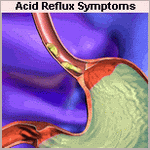
The most common complaints in patients with GERD are heartburn; regurgitation; and, occasionally, dysphagia or difficult swallowing. These represent the so-called typical symptoms of GERD. Although none of these are specific to GERD, the latter is more commonly a sign of serious underlying pathology, including esophageal carcinoma. Dysphagia should always be investigated promptly and thoroughly.
Heartburn is characterized as a substernal burning discomfort often radiating from epigastrium to sternal notch. Occasionally patients will refer to it as chest pain rather than heartburn, and the two can be difficult to distinguish. Even the location can be variable with patients occasionally experiencing discomfort in the epigastrium, base of the neck, back, or other areas. Heartburn is typically made worse by spicy foods such as tomato sauce, citrus juices, chocolate, coffee, and alcohol. It occurs 1 to 2 hours after eating, often at night and is relieved by antacids and antisecretory agents such as the over-the-counter histamine-2 blockers. It is well recognized that the severity of symptoms is not necessarily related to the severity of the underlying disease.
Regurgitation is the spontaneous return of gastric contents proximal to the GE junction. Its spontaneous nature distinguishes it from vomiting. The patient often gets a sensation that fluid or food is returning into the esophagus, even if it does not reach as high as the pharynx or mouth. It is typically worse at night in the recumbent position or when lying down after a meal. Patients commonly compensate by not eating late at night or by sleeping partially upright with several pillows or in a chair. This symptom is often less well relieved with antacids and antisecretory agents, although it may change in character from acid to a more bland nature.
Dysphagia is present in up to 40% of patients with GERD. It is generally manifested by a sensation of food hanging up in the lower esophagus (esophageal dysphagia) rather than difficulty transferring the bolus from the mouth to the esophageal inlet (oropharyngeal dysphagia). Classically dysphagia limited to only solid food, with normal passage of liquids, suggests a mechanical disorder such as a large hernia, stricture, or tumor, whereas difficulty with both solids and liquids suggest a functional or motor disorder. It often develops slowly enough that the patient may adjust his or her eating habits and not necessarily notice that it is happening. Thus, a thorough esophageal history includes an assessment of the patient's dietary history. Questions should be asked regarding the consistency of food that is typically eaten; whether the patient requires liquids with the meal; is the last to finish; has interrupted a social meal; chokes or vomits with eating; or whether he or she has been admitted on an emergency basis for food impaction. These assessments, in addition to the ability to maintain nutrition, help to quantify the dysphagia and are important in determining the indications for surgical therapy.
Many patients with GE reflux often manifest atypical symptoms, such as cough, asthma, hoarseness, and noncardiac chest pain. Atypical symptoms are the primary complaint in 20% to 25% of patients with GERD and are secondarily present in association with heartburn and regurgitation in many more. It is considerably more difficult to prove a cause-and-effect relationship between atypical symptoms and GE reflux than it is to do so for the typical symptoms. Consequently, the results of surgical therapy have been correspondingly less good. That is not to say that patients with atypical symptoms are not good candidates for antireflux surgery, because many will benefit greatly, but that in these patients it should be applied cautiously. Often a trial of high-dose proton pump inhibitors (PPIs) is helpful. Given atypical symptoms, the outcome of antireflux surgery is optimal in patients with a good response to medical treatment rather than in those who fail to respond.
The diagnosis of GERD based on symptoms alone is correct in only approximately two thirds of patients.20 This is because these symptoms are not specific for GE reflux and can be caused by other diseases such as achalasia, diffuse spasm, esophageal carcinoma, pyloric stenosis, cholelithiasis, gastritis, gastric or duodenal ulcer, and coronary artery disease. This fact underscores the need for objective diagnosis before the decision is made for surgical treatment.
Saturday, March 1, 2008
Acid Reflux (GERD) Videos
Understanding GERD (GERD #1)
What's That Pain in My Chest (Heartburn #2/GERD #2)
Treating GERD (GERD #3)
Nexium (Esomeprazole)
Prevacid (Lansoprazole)
Acid Reflux (Heartburn) Videos
Dr. Gloria Wu,"Gastroesophageal Reflux (Heartburn)" Part 2/3
Dr. Gloria Wu,"Gastroesophageal Reflux (Heartburn)" Part 3/3

