Google
 

What is acid reflux relief?

In medically oriented terms, antonyms of the word relief include pain, distress or damage. That links its meaning to both subjective and objective aspects. Subjective, denoting sensations experienced by the sufferer such as pain and objective, meaning physical findings detected by specialists which are either functional distress or organic damage. Actually relief is related to control measures and it quantitatively signifies removal of an unpleasant existence or reduction of its magnitude. The definition of relief, therefore encompasses alleviation of pain, relaxation of distress and healing of damage. Acid reflux on the other hand has two sides; the subjective side (symptoms) which reflects the symptom of heartburn and the objective side (signs) that reflects the functional and/or organic signs of esophageal changes. Acid reflux relief is therefore a broad term that covers all the measures used to control symptoms and signs of acid reflux disease. Normally, the lower esophageal sphincter remains closed except during swallowing. This prevents the passage of food and acid from the stomach into the esophagus. If the lower esophageal sphincter becomes weakened or relaxed, stomach acid may back up into the esophagus. Frequent acid reflux can irritate and inflame the lining of the esophagus, causing symptoms and signs of acid reflux. A better understanding of relief would thus entail knowledge of some aspects of normal structure and function, so that changes in the disease and its control could be easily considered. Actually acid reflux relief involves both preventive and curative measures, and in addition to treatment; orientation with the causes, symptoms and complications of acid reflux are essential for proper management. Acid reflux relief includes: dietary changes,lifestyle modifications, specific medications and surgical operations.Basic knowledge of the underlying causes and progression of acid reflux and answering frequently asked questions about its relief; add to the depth of understanding.

Sunday, November 30, 2008

Acid Reflux Relief logoRole of prokinetics in acid reflux disease


The role of prokinetic drugs in adult acid reflux disease is probably less important than in the past, due to the development of potent and safe acid suppressors. Nevertheless, prokinetic agents are still prescribed in infants and children where clinicians are more reluctant to embark in long-term acid suppression. In the future, the development of new compounds (e.g. CCK1 antagonists or GABAb agonists) that are capable of inhibiting transient lower esophageal sphincter relaxations could represent a significant progress in a field of active pharmacological research and competition.
Several prokinetic agents have been used for the management of acid reflux disease. Examples of ancillary prokinetics include: 
Bethanechol, a cholinergic agent has been shown effective in reducing symptoms and lesions of esophagitis in both adults and children. However, it also stimulates gastric acid secretion and is responsible for several side-effects. Similarly, metoclopramide (a 5HT3-antagonist), although effective on refiux symptoms at relatively large doses (at least 40 mg/day) is frequently responsible for adverse effects such as drowsiness, bowel disturbance, dizziness or even severe extrapyramidal manifestations.
Domperidone is a more recently developed dopamine antagonist related to butyrophenones that has nearly the same pharmacodynamic actions as metoclopramide on esophageal and gastric motility.
Although domperidone does not cross the blood–brain barrier and seldom causes extrapyramidal effects, it may, however, produce symptoms related to hyperprolactinaemia (galactorrhoea, or amenorrhoea). Results similar to those obtained with metoclopramide would be expected in acid reflux disease, with domperidone being better tolerated. Although metoclopramide and domperidone are still used in indigestion and other gastrointestinal motility-related disorders, they have been virtually abandoned for the treatment of acid reflux disease since the development of cisapride.
Cisapride is a prokinetic drug without an antidopaminergic effect. It is an agonist of 5-HT4 receptors and releases acetylcholine in the myenteric plexus of the gut. Cisapride increases the amplitude of esophageal body contractions, increases lower esophageal sphincter pressure (especially in reflux patients with a low tone at baseline) and accelerates gastric emptying. Nevertheless, cisapride does not change the rate of transient lower esophageal sphincter relaxations.
Although cisapride has indirect cholinomimetic effects, it does not affect gastric acid secretion. Cisapride administration (10 or 20 mg, four times a day) enhances salivary secretion in asymptomatic volunteers. This effect may contribute to esophageal clearance and benefit patients with acid reflux disease.
The efficacy of cisapride has been established beyond doubt, both in adults and children. In the short term, cisapride (10 mg four times daily or 20 mg twice daily) is more effective than placebo and equally effective as H2-receptor antagonists for symptom relief and healing of esophagitis. Large placebo-controlled trials have shown that maintenance treatment with cisapride (10 or 20 mg twice daily or 20 mg nocte) significantly reduces the 6- and 12-month relapse rate of esophagitis. The therapeutic gain, however, is mainly limited to patients with mild or moderate esophagitis.
Cisapride is usually well-tolerated, the most frequent side effects being mild diarrhoea, abdominal pain and headache. However, exceptional but lethal cardiac complications (i.e. torsades de pointes) have recently been reported. This adverse effect may reduce the role of cisapride considerably in the treatment of acid reflux disease since safe, effective, well-tolerated drugs are now available. The clinician should also be aware of the interaction between cisapride and several other drugs, such as spiramycin and ketoconazole; their concurrent use is absolutely contraindicated. The production licence for cisapride has recently been withdrawn in several countries in Europe and the USA because of life-threatening cardiac side-effects.

Saturday, September 6, 2008

Acid Reflux Relief logoProkinetic Medications

Some reports have shown that patients with acid reflux have abnormally high pyloric sphincter pressures. This has lead to the concept of “pyloric spasm” and the idea that therapies aimed at relaxing the pyloric sphincter could improve gastric emptying. Prokinetic Therapy which enhances forward gastrointestinal motility, have been proposed to address this particular pathophysiology.

Dopamine Antagonists
Metoclopramide (Reglan) is the most commonly used prokinetic drug and the agent that we employ as first line therapy. It is a central and peripheral dopamine receptor (D2) antagonist and a powerful antiemetic at the chemoreceptor trigger zone level while also being effective in improving gastric emptying by increasing antral contractions and decreasing receptive relaxation of the proximal stomach. However, as many as 40% of patients cannot tolerate metoclopramide because of central nervous system (CNS) side effects. If tolerated orally, the usual dose is 10 to 20 mg 30 minutes before meals and bedtime. One strategy for patients who do tolerate this agent orally is to also use it subcutaneously (sc) during periods of worse nausea or vomiting. Metoclopramide sc can be given by the patient (2 mL [10 mg] 2 or 3 times daily) with oral medication during and after hospitalizations while stepping down to oral therapy alone or as a bolus supplement at home to control intermittent breakthrough nausea. For patients that have intolerable side effects on metoclopromide or continued symptomatology, other agents can be used. We frequently substitute Domperidone (Motilium) for metoclopramide. It is a very effective dopamine2 antagonist, like metoclopramide, and shares its effect on upper gastrointestinal (GI) motility. It is a potent antiemetic at the chemoreceptor trigger zone but does not cross the bloodbrain barrier and, therefore, has no CNS side effects. Unfortunately, however, it is not available in the United States despite the fact that its effectiveness and safety have been established in clinical trials. Domperidone can be obtained from some compounding pharmacies in the United States and from pharmacies in other countries but is not usually covered by health insurance providers. Domperidone, at doses from 10 mg to 30 mg by mouth (po) 4 times daily (qid) (also given 30 minutes before meals and bedtime), has been shown to significantly reduce acid reflux symptoms, enhance quality of life, and accelerate gastric emptying of a solid meal. Domperidone does cause increased serum prolactin levels, and the most commonly reported side effects are gynecomastia in men and breast enlargement and lactation in women. These events occur in approximately 5% of treated patients.

Motilin Agonists
Erythromycin is a useful adjunctive therapy that can be added to metoclopramide or domperidone if symptoms persist despite maximal tolerated doses of these agents. It attaches to the motilin receptor and stimulates enteric contractility. It is not an antiemetic and concomitant agents to address nausea must also be initiated with this agent. Both intravenous and oral forms have been shown to improve gastric emptying rates, but this therapy can be complicated by cramping and abdominal pain. Low doses in a liquid (syrup) form are best to treat gastroparesis (eg, 125 mg 2 or 3 times daily). A potential concern with this agent is that it loses effectiveness over time due to down regulation of the motilin receptor.
GM611 (Mitemcinal, Chugai Pharmaceutical Company) is a macrolide that, like erythromycin, binds to the motilin receptor and stimulates enteric contractility, but does not have antimicrobial properties. It is currently in phase II trials and has been shown to increase gastric motility and gastric emptying.
Results of these treatment trials on acid reflux will be available in the near future.


5Hydroxytryptamine Agonists
Cisapride (Propulsid) is the original agent in this class and works by stimulating the stomach via 5hydroxytryptamine (5HT4) receptors. It is relatively free of CNS side effects and has been shown to improve gastric emptying of solids and liquids. Unfortunately, the unacceptably high rate of potentially fatal cardiac arrhythmias seen with this drug prompted the US Food and Drug Administration to restrict its use. Consequently, we rarely use this drug outside of special circumstances.
Mosapride (Takeda Pharmaceutical Company) is an investigational agent and is a 5HT4 agonist. It has been shown to have stimulatory effects throughout the human GI tract and has less potential for cardiac arrhythmia than its predecessor, Propulsid. It may become a useful agent in the treatment of gastroparetic patients and gastroesophageal reflux and these trials are currently being conducted.

Tegaserod (Zelnorm) (Novartis Pharmaceutical Company) is the most recently approved prokinetic agent. It has recently been FDA approved for treatment of women with constipation predominant irritable bowel syndrome (IBS). It is a partial and selective 5HT4 receptor agonist that possesses GI stimulatory effects from the esophagus to the rectum. Studies have shown
stimulatory motor effects throughout the digestive tract, and it has been shown to accelerate orocecal transit in patients with IBS. There are data showing improved gastric emptying times in patients with acid reflux and clinical trials in this subgroup of patients are ongoing. Dosing is recommended as 6 mg po twice daily but in our experience dosing up to 4 times daily is well tolerated without loose stools. This therapy is especially indicated for patients who complain of constipation in addition to their symptoms of gastroparesis. Tegaserod does not have antiemetic properties, and, therefore,would need to be given in combination with standard antiemetics.

Somatostatin (Octreotide) is used for patients with concomitant small bowel motility problems (eg, diarrhea and bacterial overgrowth). Somatostatin works by initiating a migration motor complex beginning in the small bowel, which agitates the gut, moving the bacteria further downstream and helps to sterilize the small intestine. It should be given as 50 to 100 microgram sc at night before bed. However,metronidazole or other antibiotics should be given during the day to further address bacterial overgrowth. Somatostatin actually inhibits gastric emptying and hence daytime use would require concomitant prokinetics and administration should not directly precede meals. It can be very effective for treating dumping syndrome when given preprandially in this specific clinical setting usually observed postvagotomy or gastric surgery.

Friday, July 25, 2008

Acid Reflux Relief logoAcid Reflux Diet

Typically it has been presumed that there are two mechanisms that provoke heartburn via food intake. First, some foods with a more acidic pH, such as citrus fruits, have been shown to directly irritate the lining of the esophagus and produce heartburn, and, second, some foods have
been shown to decrease the lower esophageal sphincter (LES) pressure, thereby predisposing to gastroesophageal (GE) reflux. Data with regard to the effect of a high fat meal are conlicting; however, there appears to be a consensus that chocolate does decrease the LES pressure.
Peppermint, garlic, and onions are food constituents that clinically appear to induce heartburn
in many patients. These compounds have a similar derivation and are often categorized as carminatives. In a clinical study to directly test one of these constituents (ie, raw onions), and its effect on postprandial reflux. In this study, individuals had to eat a hamburger with and without a large raw onion. In the onion condition it was shown that there was a significant postprandial increase in esophageal acid contact. Peppermint has also been shown to induce GE reflux, but this may be confounded by the fact that dissolving a peppermint may have the salubrious effect of enhancing salivation, which will facilitate acid clearance. Chewing gum may also induce increased salvation with similar effects on esophageal acid clearance.
Heartburn and esophageal irritation are common experiences in individuals with an acid sensitive esophagus who ingest citrus fruit juices. A recently published survey showed a significant correlation between the acidity of the citrus juices and heartburn score.
On this basis, it would seem reasonable to advise patients to avoid chocolate, citrus fruits, and fruit juices, as well as carminatives, such as onions and garlic.
Data with regard to high fat content on LES pressure are equivocal, but it would seem rational to advise individuals to avoid high fat foods, at least with regard to their effect on delayed gastric emptying and GE reflux.
Carbonation and caffeine.
There's no differences between carbonated water, caffeine-free Pepsi, or regular Pepsi, the LES changes are due to gas rather than caffeine level or pH. The difference between regular coffee and decaffeinated coffee, It has been found that decaffeinated coffee indeed did reduce the per-
cent of acid contact time in the esophagus.
Gastric Distention
An obvious and inexorable effect of eating is gastric distention. Because evidence is now clear that gastric distention does induce transient lower esophageal sphincter relaxations (TLESR), it would seem that food ingestion alone would predispose to reflux via the mechanism of TLESR. Again, although no randomized control trial exists to document the efficacy of this measure, it would seem prudent to advise patients to avoid large volume meals as well as carbonation.

Monday, July 7, 2008

Acid Reflux Relief logoEffective Acid Reflux Relief

Treatment of Acid Reflux Disease is primarily medical, the mainstays being lifestyle modifications and drug therapy. The goals of treatment are to relieve symptoms and prevent relapse and complications. All patients should be advised about lifestyle modifications that help reduce symptoms and prevent relapse. Antacids or antacid-alginate combinations are recommended for safe, prompt, inexpensive relief of heartburn. The same agents, however, are poorly suited for regular use because of poor palatability and durability and side effects such as diarrhea, constipation, and possible magnesium or aluminum toxicity in renal patients. Protection against recurrence of heartburn is provided by acid-suppressing medications such as H2-receptor antagonists and PPIs. H2-receptor antagonists reduce gastric acid secretion moderately by inhibiting one of three acid-stimulating receptors on the basolateral membrane of the parietal cell. When prescribed twice a day, they can control symptoms in about 50% of Acid Reflux Disease patients and heal erosions in about 30%. PPIs irreversibly inhibit the H+, K+-ATPase or proton pump, the final common pathway for acid secretion on the apical membrane of the parietal cell. Consequently, PPIs markedly reduce gastric acidity with once-a-day dosing and provide relief of symptoms and healing of lesions in about 80 to 90% of Acid Reflux Disease patients. H2-receptor antagonists (+30 years) and PPIs (≈15 years) have excellent safety profiles. PPI safety beyond 15 years remains unclear because of uncertainty about the long-term risk for chronic gastric hypoacidity and hypergastrinemia. Although vitamin B12 levels can be reduced with chronic PPI use, clinically significant vitamin B12 deficiency has not been reported, so an increase in vitamin B12 intake is not currently recommended.

Early endoscopy is indicated for those with alarm symptoms. Endoscopy is also indicated for patients who fail once-a-day PPI therapy to confirm the diagnosis and assess severity, including the presence of Barrett's esophagus (see later). Testing for H. pylori is not recommended because the organism is not etiologic in Acid Reflux Disease and, when eradicated, may make treatment more difficult.

Failures with once-a-day PPI therapy are treated with twice-daily PPI therapy with or without H2-receptor antagonists at bedtime for 6 to 8 weeks, and patients who fail this regimen undergo esophageal pH monitoring during therapy to assess for control of esophageal acidity. If the acidity is controlled, the symptoms are not mediated by acid.

Effective therapy is often accompanied by relapse when medication ceases, especially in patients with erosive esophagitis, in whom maintenance therapy is indicated. Patients requiring maintenance therapy should undergo at least one endoscopy procedure to determine whether Barrett's esophagus is present. If endoscopy reveals NERD, no further endoscopy is necessary and treatment is guided by symptoms. If endoscopy reveals erosive esophagitis, treatment to healing should be documented by endoscopy so that Barrett's esophagus can be effectively established or excluded. Once Barrett's esophagus is excluded, endoscopy is unnecessary and treatment is guided by symptoms because subsequent relapse and treatment will rarely result in Barrett's esophagus.

Acid Reflux Relief

Acid Reflux Relief

Acid Reflux Relief

Saturday, June 21, 2008

Acid Reflux Relief logoTREATMENT OF ACID REFLUX COMPLICATIONS

TREATMENT OF ACID REFLUX COMPLICATIONS
Strictures
Mildly symptomatic esophageal strictures can be handled by careful attention to dietary intake, and use of medical therapy, primarily proton-pump inhibitors. Short, simple strictures can be dilated with weighted rubber or Teflon dilators (e.g., Hurst-Maloney). Tortuous or angulated strictures are more easily approached over a previously placed guidewire passed through an endoscope or under radiographic control (Savary dilators). Graded-steel olives (Eder-Puestow olive dilators), a dilator with graded increases of size (Celestin dilator), or a balloon with a fixed maximal diameter (Cooke balloon) can be passed over the previously placed wire. Alternatively, a balloon of fixed maximal diameter can be passed through the large channel of an endoscope during diagnostic endoscopy and dilated under direct vision (through-the-scope [TTS] dilation). Once the lumen is restored to a diameter of 13 to 15 mm, most patients swallow without difficulty. If the stricture is stable and requires dilation only every 4 to 6 months, no other therapy is necessary.
High-dose H2 -antagonists or, preferably, proton-pump inhibitors and dilation of the stricture can lead to healing of the mucosa and less need for repeated stricture dilation. Patients who do not tolerate dilation or require vigorous dilation every 3 to 4 weeks need a definitive antireflux operation, following which the stricture may regress. If strictures persist after antireflux surgery, esophageal replacement by colon, jejunum, or stomach is a surgical maneuver of last resort associated with a relatively high morbidity and mortality. Patients afflicted by strictures may have significant lung and cardiovascular disease that makes them unsuitable operative candidates.
Ulcers
Esophageal ulcers also represent a major therapeutic problem. They usually require treatment with a proton-pump inhibitor.
Barrett's Esophagus
Barrett's (columnar) epithelium may be premalignant and can be removed only by esophageal resection. Adequate antireflux therapy with high-dose H2 -antagonists or with a proton-pump inhibitor causes regression of columnar epithelium in some patients.
Patients with Barrett's epithelium should be followed up with periodic endoscopic biopsies every 1 to 3 years to look for dysplasia and early changes of adenocarcinoma. The persistence of confirmed high-grade dysplasia is an indication for esophagectomy, because high-grade dysplasia may progress to carcinoma and because coexistent carcinoma may be undetected on biopsy. If low-grade dysplasia is present, the patient is treated medically with proton-pump inhibitors and undergoes biopsy every 6 to 12 months. Experimental endoscopic ablation therapies using photodynamic therapy, laser, or multipolar electrocoagulation are being tried to remove the columnar epithelium with the hope of subsequent growth of the normal squamous epithelium, primarily in patients with low-grade dysplasia or in patients with high-grade dysplasia who are not surgical candidates. Following these new ablation techniques, either long-term gastric acid suppression or laparoscopic Nissen fundoplication is needed to control the reflux and prevent recurrence of Barrett's epithelium.
Pulmonary Complications
Treatment of the pulmonary complications of reflux in adults relies on improved night posture, gastric acid suppressants, and prokinetic agents. Caution is advised before recommending esophageal surgery in patients with reflux and predominant pulmonary problems, because the cause-and-effect relationship may be uncertain in individual patients.

Tuesday, June 10, 2008

Acid Reflux Relief logoBarrett's esophagus Treatment: Photodynamic therapy

Photodynamic therapy may effectively treat superficial esophageal cancer and Barrett's esophagus. The treatment begins with an injection of a light sensitive medication. Your normal body cells can get rid of this medication, but precancerous and cancerous cells in your esophagus cannot, so it accumulates in these tissues and makes them sensitive to light. Two days after the injection, your doctor inserts an endoscope — a long, flexible instrument — into the diseased area of your esophagus. The light from a laser on the endoscope activates the medication, killing the light sensitive cancerous cells.

This medication will make all the cells in your body light sensitive, so talk to your doctor about how you can avoid sunlight for up to 6 weeks after the treatment.

Saturday, June 7, 2008

Acid Reflux Relief logoHeartburn Relief : Acid Reflux Treatment

The management of frequent heartburn entails certain measures to confirm the diagnosis of GERD. Following these diagnostic procedures, treatment of acid reflux disease should be considered.
Acid Reflux Treatment

The aim of treatment are to reduce refluxing, render the refluxate harmless, improve esophageal clearance, and protect the esophageal mucosa. The management of non-complicated cases generally includes weight reduction, sleeping with the head of the bed elevated by about 4 to 6 in. with blocks, and elimination of factors that increase abdominal pressure. Patients should not smoke and should avoid consuming fatty foods, coffee, chocolate, alcohol, mint, orange juice, and certain medications (such as anticholinergic drugs, calcium channel blockers, and other smooth-muscle relaxants). They should also avoid ingesting large quantities of fluids with meals. In mild cases, life-style changes and over-the-counter antisecretory agents may be adequate. In moderate cases, H2receptor blocking agents (cimetidine, 300 mg; ranitidine, 150 mg bid; famotidine, 20 mg bid; nizatidine 150 mg bid) for 6 to 12 weeks are effective in symptom relief. Higher doses are necessary for healing erosive esophagitis, but proton pump inhibitors (PPIs) are more effective in this setting.

In cases resistant to H2receptor blockers and severe cases, rigorous acid suppression with aPPI is recommended. The PPIs are comparably effective: omeprazole (40 mg/d), lansoprazole (30 mg/d), pantoprazole (40 mg/d), and rabeprazole (20 mg/d) for 8 weeks can heal erosive esophagitis in up to 90% of patients. Reflux esophagitis requires prolonged therapy, for 3 to 6 months or longer if the disease recurs quickly. After initial therapy, a lower maintenance dose of PPI is used. Side effects are minimal. Aggressive acid suppression causes hypergastrinemia but does not increase the risk for carcinoid tumors or gastrinomas. Vitamin B12 absorption is compromised by the treatment. Patients with reflux esophagitis who have complications, such as Barrett's esophagus with concomitant esophagitis, should be treated vigorously. Patients who have an associated peptic stricture are treated with dilators to relieve dysphagia as well as provided with vigorous treatment for reflux.

Antireflux surgery, in which the gastric fundus is wrapped around the esophagus (fundoplication), increases theLESpressure and should be considered for patients with resistant and complicated reflux esophagitis that does not respond fully to medical therapy or for patients for whom long-term medical therapy is not desirable. Laparoscopic fundoplication is the surgery of choice. Ideal candidates for fundoplication are those in whom motility studies show persistently inadequate LES pressure but normal peristaltic contractions in the esophageal body.

Patients with alkaline esophagitis are treated with general antireflux measures and neutralization of bile salts with cholestyramine, aluminum hydroxide, or sucralfate. Sucralfate is particularly useful in these cases, as it also serves as a mucosal protector.

Thursday, June 5, 2008

Acid Reflux Relief logoAcid Reflux Relief : Combination Therapy

Most patients treated with PPIs in conventional dosages do not exhibit complete suppression of stomach acid secretion. Approximately 70% of individuals who take a PPI twice a day experience nocturnal stomach acid breakthrough (defined as a stomach pH lesser than 4 for more than 1 hour at night). Brief episodes of acid reflux occur frequently during these breakthrough periods in patients with GERD. For some patients taking a PPI twice daily, nocturnal acid breakthrough can be abolished by adding a histamine H2-receptor blocker at bedtime. It is not clear that this approach is desirable, however. Complete elimination of acid reflux usually is not necessary to effect the healing of reflux esophagitis. Indeed, most patients who are treated with a PPI in conventional dosage exhibit complete healing of their symptoms and signs of GERD. No clear clinical benefit yet has been demonstrated for the practice of adding a histamine H2-receptor blocker at bedtime to PPI therapy.

A few older investigations have explored the value of combination drug therapy for the healing of GERD. The great efficacy of the PPIs used as single agents in this condition has discouraged investigators from undertaking new studies on combination therapy. Drug combinations that have been studied have included an H2 blocker plus either sucralfate or a prokinetic agent. Cimetidine (1200 mg/d) combined with sucralfate (5 g/d) was found to be superior to cimetidine alone for relieving daytime heartburn and for improving the endoscopic signs of esophagitis. For patients unresponsive to treatment with cimetidine alone, the addition of metoclopramide resulted in symptomatic improvement significantly more often than the addition of placebo, but side effects of metoclopramide were frequent. A combination of ranitidine (300 mg/d) plus metoclopramide (40 mg/d) was not found to be as effective as omeprazole alone (20 mg/d) in healing the signs and symptoms of esophagitis. Some studies explored combination therapy with the prokinetic agent cisapride, but these studies are of historical interest only because cisapride has been withdrawn from general use due to serious side effects (lethal arryhythmias). For patients with moderately severe reflux esophagitis, the use of combination therapy may eliminate the need for treatment with a PPI. However, the addition of a second medication increases the cost of therapy and the potential for side effects. Furthermore, the long-term benefit of combination therapy has not been demonstrated. For patients who are refractory to single-agent therapy (with an H2 blocker, sucralfate, or a prokinetic), a change to a PPI generally is more likely to effect healing than the addition of a second drug.

Friday, May 23, 2008

Acid Reflux Relief logoTreatment of Barrett's esophagus

The relief of symptoms remains the primary force driving antireflux surgery in patients with Barrett's esophagus. Healing of esophageal mucosal injury and the prevention of disease progression are important secondary goals. In this regard, patients with Barrett's esophagus are no different than the broader population of patients with GE reflux. Antireflux surgery should be considered when patient factors suggest severe disease or predict the need for long-term medical management, both of which are almost always true in patients with Barrett's esophagus.
PPI therapy, both to relieve symptoms and to control any coexistent esophagitis or stricture, is an acceptable treatment option in patients with Barrett's esophagus. Once initiated, however, most patients with Barrett's will require lifelong treatment. Complete control of reflux with PPI therapy can be difficult, however, as has been highlighted by studies of acid breakthrough while on therapy. Ablation trials have shown that mean doses of 56 mg of omeprazole are necessary to normalize 24-hour esophageal pH studies. Antireflux surgery likely results in more reproducible and reliable elimination of reflux of both acid and duodenal content, although long-term outcome studies suggest that as many as 25% of patients postfundoplication have persistent pathologic esophageal acid exposure confirmed by 24-hour pH studies.58
An important consideration is that patients with Barrett's esophagus generally have severe GERD, with its attendant sequelae such as large hiatal hernia, stricture, shortened esophagus, and poor motility. These anatomic and physiologic features make successful antireflux surgery a particular challenge in this population. Indeed, recent data suggest that antireflux surgery in patients with Barrett's esophagus may not be as successful in the long term as in those without Barrett's.
Studies focusing on the symptomatic outcome following antireflux surgery in patients with Barrett's esophagus document excellent to good results in 72% to 95% of patients at 5 years following surgery The outcome of laparoscopic Nissen fundoplication in patients with Barrett's esophagus has been assessed at 1 to 3 years after surgery.Reflux symptoms were absent postoperatively in 79% of the patients. Postoperative 24-hour pH was normal in 17 of 21 (81%) patients. Ninety-nine percent of the patients considered themselves cured or improved, and 97% were satisfied with the surgery.

Tuesday, May 20, 2008

Acid Reflux Relief logoWhat is Barrett's esophagus?

Barrett's esophagus is the metaplastic complication of Acid Reflux Disease.
It is the condition whereby the tubular esophagus is lined with columnar epithelium rather than squamous epithelium was first described by Norman Barrett in 1950. He incorrectly believed it to be congenital in origin. It is now realized that it is an acquired abnormality, occurring in 7% to 10% of patients with GERD, and represents the end stage of the natural history of this disease. It is also understood to be distinctly different from the congenital condition in which islands of mature gastric columnar epithelium are found in the upper half of the esophagus.
The definition of Barrett's esophagus has evolved considerably over the past decade. Traditionally, Barrett's esophagus was identified by the presence of any columnar mucosa extending at least 3 cm into the esophagus. Recent data indicating that specialized intestinal-type epithelium is the only tissue predisposed to malignant degeneration, coupled with the finding of a similar risk of malignancy in segments of intestinal metaplasia less than 3 cm long, have resulted in the diagnosis of Barrett's esophagus, given any length of endoscopically visible tissue that is intestinal metaplasia on histology. Whether to call long segments of columnar mucosa without intestinal metaplasia Barrett's esophagus is unclear. The hallmark of intestinal metaplasia is the presence of goblet cells. Recent studies have identified a high prevalence of biopsy-proved intestinal metaplasia at the cardia, in the absence of endoscopic evidence of a columnar-lined esophagus. The significance and natural history of this finding remains unknown. The term Barrett's esophagus should currently be used in the setting of an endoscopically visible segment of intestinal metaplasia of any length or columnar replacement of the esophagus of 3 cm or more.
Factors predisposing to the development of Barrett's esophagus include early-onset GERD, abnormal LES and esophageal body physiology, and mixed reflux of gastric and duodenal contents into the esophagus. Direct measurement of esophageal bilirubin exposure as a marker for duodenal juice has shown that 58% of the patients with GERD have increased esophageal exposure to duodenal juice and that this exposure is most dramatically related to Barrett's esophagus.
Pathophysiology of Barrett's Metaplasia
Recent studies suggest that the metaplastic process at the GE junction may begin by conversion of distal esophageal squamous mucosa to cardiac-type epithelium, heretofore presumed to be a normal finding. This is likely due to exposure of the distal esophagus to excess acid and gastric contents via prolapse of esophageal squamous mucosa into the gastric environment. This results in inflammatory changes at the GE junction or a metaplastic process, both of which may result in the loss of muscle function and a mechanically defective sphincter allowing free reflux with progressively higher degrees of mucosal injury. Intestinal metaplasia within the sphincter may result, as in Barrett's metaplasia of the esophageal body. This mechanism is supported by the finding that as the severity of GERD progresses, the length of columnar lining above the anatomic GE junction is increased.

Friday, May 16, 2008

Acid Reflux Relief logoAcid Reflux Disease Complications

The complications of GE reflux result from the damage inflicted by gastric juice on the esophageal mucosa. Mucosal complications, include esophagitis and stricture. The prevalence and severity of complications is related to the degree of loss of the GE barrier, defects in esophageal clearance, and the content of refluxed gastric juice.
The potential injurious components that reflux into the esophagus include gastric secretions, such as acid and pepsin, biliary and pancreatic secretions that regurgitate from the duodenum into the stomach, and toxic compounds generated in the mouth, esophagus, and stomach by the action of bacteria on dietary substances.
Studies have shown that acid alone does minimal damage to the esophageal mucosa, but the combination of acid and pepsin is highly deleterious. Hydrogen ion injury to the esophageal squamous mucosa occurs only at a pH below 2. In acid refluxate, the enzyme pepsin appears to be the major injurious agent. Similarly, the reflux of duodenal juice alone does little damage to the mucosa, whereas the combination of duodenal juice and gastric acid is particularly noxious. Reflux of bile and pancreatic enzymes into the stomach can either protect against or augment esophageal mucosal injury. For instance, the reflux of duodenal contents into the stomach may prevent the development of peptic esophagitis in a patient whose gastric acid secretion maintains an acid environment, because the bile salts would attenuate the injurious effect of pepsin and the acid would inactivate the trypsin. Such a patient would have bile-containing acid gastric juice that, when refluxed, would irritate
the esophageal mucosa but cause less esophagitis than if it were acid gastric juice containing pepsin. In contrast, the reflux of duodenal contents into the stomach of a patient with limited gastric acid secretion can result in esophagitis, because the alkaline intragastric environment would support optimal trypsin activity, and the soluble bile salts with a high pKa would potentiate the enzyme's effect. Hence, duodenal-gastric reflux and the acid secretory capacity of the stomach interrelate by altering the pH and enzymatic activity of the refluxed gastric juice to modulate the injurious effects of enzymes on the esophageal mucosa.
This disparity in injury caused by acid and bile alone, as opposed to the gross esophagitis caused by pepsin and trypsin, provides an explanation for the poor correlation between the symptom of heartburn and endoscopic esophagitis. The reflux of acid gastric juice contaminated with duodenal contents could break the esophageal mucosal barrier, irritate nerve endings in the papillae close to the luminal surface, and cause severe heartburn. Despite the presence of intense heartburn, the bile salts present would inhibit pepsin, the acid pH would inactivate trypsin, and the patient would have little or no gross evidence of esophagitis. In contrast, the patient who refluxed alkaline gastric juice may have minimal heartburn because of the absence of hydrogen ions in the refluxate but have endoscopic esophagitis because of the bile salt potentiation of trypsin activity on the esophageal mucosa. This is supported by recent clinical studies which indicate that the presence of alkaline reflux is associated with the development of mucosal injury.
Although numerous studies have suggested the reflux of duodenal contents into the esophagus in patients with GERD, few have measured this directly. The components of duodenal juice thought to be most damaging are the bile acids and, as such, they have been the most commonly studied. Most studies shown that, patients with GERD have greater and more concentrated bile acid exposure to the esophageal mucosa than do normal subjects. This increased exposure occurs most commonly during the supine period while asleep and during the upright period following meals. Most studies have identified the glycine conjugates of cholic, deoxycholic, and chenodeoxycholic acids as the predominant bile acids aspirated from the esophagus of patients with GERD, although appreciable amounts of taurine conjugates of these bile acids were also found. Other bile salts were identified but in small concentrations. This is as one would expect because glycine conjugates are three times more prevalent than taurine conjugates in normal human bile.
The potentially injurious action of toxic compounds either ingested or newly formed on the mucosa of the GE junction and distal esophagus has long been postulated. Investigators have recently shown that dietary nitrate consumed in the form of green vegetables and food contaminated by nitrate-containing fertilizers results in the generation of nitric oxide at the GE junction in concentrations high enough to be potentially mutagenic. Previous studies have shown that nitrate ingested in food is reabsorbed in the small bowel with approximately 25% resecreted into the mouth via the salivary glands. Oral bacteria chemically transform the relatively innocuous nitrate to the more toxic nitrite, which is swallowed and subsequently converted to nitric oxide and other toxic nitroso-compounds by acid and ascorbic acid in the stomach. Whether this mechanism in fact contributes to injury and or neoplastic transformation in the upper stomach, GE junction, and distal esophagus is currently unknown.

Tuesday, May 13, 2008

Acid Reflux Relief logoAnti reflux Surgery Complications

Postoperative complications were found to occur in approximately 8% of patients, with the rate of conversion to an open procedure of about 4%. The most common perioperative complication was early wrap herniation (1.3%), defined as occurring within 48 hours of surgery. This is one complication that may be more common with the laparoscopic than open approach. The explanation for this is unclear but may be related to the opening of tissue planes by the pneumoperitoneum and the reduced tendency for adhesion formation after laparoscopic compared to open surgery. In an attempt to eliminate this complication, most surgeons routinely perform a crural repair.
Both pneumothorax and pneumomediastinum have been reported. The occurrence of pneumothorax is related to breach of either pleural membrane, usually the left, during the hiatal dissection. Chest drain insertion is usually not required because accumulated carbon dioxide rapidly dissipates following release of pneumoperitoneum by a combination of positive pressure ventilation and absorption.
As with any laparoscopic procedure, instrumental perforation of the hollow viscera may occur. Early esophageal perforation may arise during passage of the bougie, during the retroesophageal dissection, or during suture pull-through. Late esophageal perforation is related to diathermy injury at the time of mobilization. Gastric perforations usually resulted from excessive traction on the fundus for retraction purposes. Recognition of the problem at the time of surgery requires repair, which may be performed either laparoscopically or by an open technique.
Hemorrhage during the course of laparoscopic fundoplication usually arises from the short gastric vessels or spleen. Rarer causes include retractor trauma to the liver, injury to the left inferior phrenic vein, an aberrant left hepatic vein, or the inferior vena cava. Cardiac tamponade as a result of right ventricular trauma has also been reported. Major vascular injury mandates immediate conversion to an open procedure to achieve hemostasis.

Acid Reflux Relief logoAcid reflux disease facts

The diagnosis of GE reflux based on symptoms alone is correct in only approximately two thirds of patients because the symptoms are often nonspecific and can be caused by other conditions.

The three characteristics of the LES that maintains its resistance or barrier function to intragastric and intraabdominal pressure challenges are pressure, overall length, and length exposed to the positive pressure environment of the abdomen.

An important complication of GE reflux is the development of reflux-induced respiratory symptoms either with or without heartburn.

Antireflux surgery improves respiratory symptoms in nearly 90% of children and 70% of adults with asthma and reflux disease.

Factors predisposing to the development of Barrett's esophagus include early-onset GERD, abnormal lower esophageal and esophageal body physiology, and mixed reflux of gastric and duodenal contents into the esophagus.

PPI therapy, both to relieve symptoms and to control esophagitis, is an acceptable treatment, although most patients will require life-long treatment.

Progression of nondysplastic Barrett's epithelium occurs with 5% to 10% of patients per year progressing to dysplasia and 0.5% to 1% per year progressing to cancer

The standard of care of the treatment of confirmed Barrett's esophagus with high-grade dysplasia is esophagectomy because approximately 50% will harbor invasive cancer

Three factors predictive of a successful outcome following antireflux surgery are (a) an abnormal score on 24-hour esophageal pH monitoring; (b) the presence of typical symptoms of GERD, namely heartburn or regurgitation; and (c) symptomatic improvement in response to acid-suppression therapy prior to surgery.

Saturday, May 3, 2008

Acid Reflux Relief logoEndoscopic investigation of gastro-esophageal reflux

The development of fibre-optic endoscopy revolutionized the ability to investigate the gastrointestinal tract. Flexible endoscopes often have a diameter of less than 1 cm, with a control head and a flexible shaft with a manoeuvrable tip. The head is connected to a light source and can transmit images to a video image screen.
All endoscopes have multiple small lumens allowing transmission of air and water and for suction. The suction channel can also be used for the passage of interventional devices, for example biopsy forceps. The ability to transmit air (insufflation) allows the endoscopist to inflate the lumen to obtain optimal views. The water channel provides a means of washing mucosal surfaces, and suction maybe used to remove pools of fluid within the gastrointestinal tract, thus ensuring that all mucosal surfaces are inspected.
Diagnostic indications include the investigation of gastro-esophageal reflux (diagnostic investigation or surveillance endoscopy of Barrett's esophagus).
Therapeutic procedures that can be undertaken include mucosal biopsy, stent insertion for strictures, and dilatation of strictures.
Patient preparation
Informed consent is required, and patients are fasted for 4-6 hours prior to the procedure. Although most patients do not require sedation, the choice is offered and discussed. Sedation involves the use of a short-acting benzodiazepine which provides a sedative and amnesic effect. Monitoringis required (pulse oximetry) with sedation due to the risk of respiratory depression. Antibiotic prophylaxis is administered to patients with heart valve disease to prevent bacterial endocarditis.
Procedure
A mouthguard is used. The endoscope is introduced into the pharynx, then the oesophagus. Patients may retch during this procedure. The endoscope is progressively introduced to inspect the oesophagus, stomach and proximal small bowel (duodenum).
Complications
The overall complication rate is approximately 1 per 1000 with a mortality rate of approximately 1 per 25000. Complications include bleeding, perforation and respiratory arrest (a complication of sedation).
Post procedure care
Patients are monitored in a recovery area until safe for discharge.

Friday, April 18, 2008

Acid Reflux Relief logoHiatal hernia and GERD

A hiatal hernia is the protrusion (or herniation) of the upper part of the stomach into the thorax through a tear or weakness in the diaphragm. The most common (95%) is the sliding hiatal hernia, where the gastroesophageal junction moves above the diaphragm together with some of the stomach.

Hiatal hernias affect anywhere from 1 to 20% of the population. Of these, 9% are symptomatic, depending on the competence of the lower esophageal sphincter (LES). 95% of these are "sliding" hiatal hernias, in which the LES protrudes above the diaphragm along with the stomach, and only 5% are the "rolling" type (paraesophageal), in which the LES remains stationary but the stomach protrudes above the diaphragm. People of all ages can get this condition, but it is more common in older people.

There are three anatomical factors that contribute to the prevention of reflux: (i) the acute angle at which the esophagus enters the stomach with its associated valve (called the angle of His); (ii) the phrenoesophageal ligament that seems to hold the gastroesophageal junction in place; and (iii) the diaphragmatic crura (which act to prevent reflux due to the position of the stomach below the diaphragm) and the intra-abdominal segment of lower esophageal sphincter. It has been reported that the angle of His is larger in the symptomatic group of endoscopically diagnosed acid reflux than in the asymptomatic group, indicating the importance of this angle with regard to gastroesophageal reflux.
Following any disturbance at any of these mechanisms, a sliding hernia would occur and cause gastroesophageal reflux. It is presumed that the tissues around the diaphragmatic hiatal that prevent GER maintain the form of cardia whenever abdominal pressure is increased.

The following are possible causes or contributing factors for having a hiatal hernia.
* Increased pressure within the abdomen caused by:
o Heavy lifting or frequent bending over
o Frequent or hard coughing
o Hard sneezing
o Violent vomiting
o Straining with constipation
o Obesity (extra weight pushes down on the abdomen increasing the pressure)
o Use of the sitting position for defecation.
* Heredity
* Smoking
* Drug use, such as cocaine.
* Stress

If mechanical forces set in play by stomach distention are important in pulling on the terminal esophagus and shortening the length of the high-pressure zone or sphincter, then the geometry of the cardia, that is, the presence of a normal acute angle of His or the abnormal dome architecture of a sliding hiatal hernia, should influence the ease with which the sphincter is pulled open. A close relationship exists between the degree of stomach distention necessary to overcome the high-pressure zone and the morphology of the cardia. Greater stomach dilatation, as reflected by a higher pressure inside stomach, is necessary to open the sphincter in patients with an intact angle of His compared to those with a hiatal hernia. This is what would be expected if the high-pressure zone were shortened by mechanical forces and accounts for why a hiatal hernia is often associated with the presence of GERD.

The symptoms include acid reflux, and pain, similar to heartburn, in the chest and upper stomach.
In most patients, hiatal hernias cause no symptoms. Sometimes patients experience heartburn and regurgitation, when stomach acid refluxes back into the esophagus.
A hiatal hernia per se may not cause any symptoms. The condition promotes reflux of stomach contents (via its direct and indirect actions on the anti-reflux mechanism) and thus is associated with gastroesophageal reflux disease (GERD). In this way a hiatal hernia is associated with all the potential consequences of GERD - heartburn, esophagitis, Barrett's esophagus and esophageal cancer. However the risk attributable to the hiatal hernia is difficult to quantify, and at most is low.

Besides discomfort from GERD and dysphagia, hiatal hernias can have severe consequences for patients if not treated. While sliding hernias are primarily associated with gastroesophageal acid reflux, rolling hernias can strangulate a portion of the stomach above the diaphragm. This strangulation can result in esophageal or GI tract obstruction and the tissue even become ischemic and necrose.
Another severe complication, although very rare, is a large herniation that can restrict the inflation of a lung, causing pain and breathing problems.

The diagnosis of a hiatal hernia is typically made through an upper GI series or endoscopy.
Gastroesophageal scintigraphy is a sensitive and non-invasive method for evaluation of GER. Most patients with typical GER symptoms exhibit scintigraphic evidence of reflux, and the severity of their reflux symptoms is correlated with the scintigraphic reflux index. The reflux index also increases as endoscopic esophagitis become more severe. The severity of reflux symptoms could be correlated with the reflux index, and the index increased significantly as endoscopic esophagitis became more severe. Therefore, the reflux index appeared to be a reliable measure of GER.
The severity of sliding hernia could be classified by esophagogastroscopy. Using the diameter of the fiberscope (9–10 mm) to estimate hernia size; also on the basis of severity. Evaluation of the relationship between the reflux index and the endoscopic grade of hiatal hernia demonstrated that the index was significantly higher in the mild hernia group compared with the severe group. Endoscopic esophagitis was found to be more prevalent in the severe group than in the mild group.
The relationship between reflux esophagitis and the size of the sliding hernia was also studied, concluding that hernia was significantly larger in patients with erosive esophagitis than in those without esophagitis and that hiatal hernia size was a reliable indicator of the severity of esophagitis.

In most cases, sufferers experience no discomfort and no treatment is required. However, when the hiatal hernia is large, or is of the paraesophageal type, it is likely to cause esophageal stricture and discomfort. Symptomatic patients should elevate the head of their beds and avoid lying down directly after meals until treatment is rendered. If the condition has been brought on by stress, stress reduction techniques may be prescribed, or if overweight, weight loss may be indicated. Medications that lower the lower esophageal sphincter (or LES) pressure should be avoided. Antisecretory drugs like proton pump inhibitors and H2 receptor blockers can be used to reduce acid secretion.
Where hernia symptoms are severe and chronic acid reflux is involved, surgery is sometimes recommended, as chronic reflux can severely injure the esophagus and even lead to esophageal cancer.
The surgical procedure used is called Nissen fundoplication. In fundoplication, the stomach fundus (upper part) of the stomach is wrapped, or plicated, around the inferior part of the esophagus, preventing herniation of the stomach through the hiatus of the diaphragm and the reflux of stomach acid. The procedure is now commonly performed laparoscopically. With proper patient selection, laparoscopic fundoplication has low complication rates and a quick recovery.
Complications include gas bloat syndrome, dysphagia (difficult swallowing), dumping syndrome, excessive scarring, and rarely, achalasia. The procedure sometimes fails over time, requiring a second surgery to make repairs.

Thursday, March 20, 2008

Acid Reflux Relief logoGERD Asthma Treatment

Once GERD is suspected or thought to be responsible for asthma symptoms, treatment may be with either prolonged PPI (proton pump inhibitor) therapy or anti reflux surgery. A 3- to 6-month trial of high-dose PPI therapy [twice a day (b.i.d.) or three times a day (t.i.d.) dosing] may help confirm (by virtue of symptom resolution) that GERD is partly or completely responsible for the asthma symptoms. The persistence of symptoms despite PPI treatment, however, does not necessarily rule out GERD as a potential contributor.
Based on reported observations, relief of asthma symptoms can be anticipated for 25% to 50% of patients with GERD asthma treated with anti reflux medications.
Fewer than 15%, however, can be expected to have objective improvements in their pulmonary function. The reason for this apparent paradox may be that most studies employed relatively short courses of anti reflux therapy (less than 3 months). This time period may have been sufficient for symptomatic improvement but insufficient for recovery of pulmonary function. The chances of success with medical treatment are likely directly related to the extent of GERD elimination. The conflicting findings of reports of anti reflux therapy may well be to the result of inadequate control of GERD in some studies. The literature indicates that anti reflux surgery improves asthma symptoms in nearly 90% of children and 70% of adults with asthma and GERD. Improvements in pulmonary function were demonstrated in around one third of patients. Comparison of the results of uncontrolled studies of each form of therapy and the evidence from the two randomized controlled trials of medical versus surgical therapy indicate that fundoplication is the most effective therapy for GERD asthma. The superiority of the surgical anti reflux barrier over medical therapy is probably most noticeable in the supine posture, which corresponds with the period of acid breakthrough with PPI therapy and is the time in the circadian cycle when asthma symptoms and peak expiratory flow rates are at their worst.
It is also important to realize that, in asthmatic patients with a non reflux induced motility abnormality of the esophageal body, performing an anti reflux operation may not prevent the aspiration of orally regurgitated, swallowed liquid or food. This can result in asthma symptoms and airway irritation that may elicit an asthmatic reaction. This factor may explain why surgical results appear to be better in children than adults, because disturbance of esophageal body motility is more likely in adult patients.

Monday, March 17, 2008

Acid Reflux Relief logoGERD Asthma

Two mechanisms have been proposed as the pathogenesis of GERD asthma. The first, the so-called reflux theory, maintains that asthma is the result of the aspiration of gastric contents. The second or reflex theory maintains that vagally mediated bronchoconstriction follows acidification of the lower esophagus. The evidence supporting a reflux mechanism is fivefold. First, clinical studies have documented a strong correlation between idiopathic pulmonary fibrosis and hiatal hernia. The presence of GERD was shown to be highly associated with several pulmonary diseases, not only asthma, in recent studies. Second, pathologic acid exposure in the proximal esophagus is often identified in patients with asthma and reflux disease. Third, scintigraphic studies have shown aspiration of ingested radioisotope in some patients with reflux and respiratory symptoms. Fourth, simultaneous tracheal and esophageal pH monitoring in patients with reflux disease has documented tracheal acidification in concert with esophageal acidification. Finally, animal studies have shown that tracheal instillation of hydrochloric acid profoundly increases airways resistance.
A reflex mechanism is primarily supported by the fact that bronchoconstriction occurs following the infusion of acid into the lower esophagus. This can be explained by the common embryologic origin of the tracheoesophageal tract and its shared vagal innervation. Second, patients with asthma and pathologic distal esophageal acid exposure but normal proximal esophageal acid exposure may experience an improvement in their asthma after antireflux therapy.
The primary challenge in implementing treatment for reflux-associated asthma lies in establishing the diagnosis. In patients with predominantly typical reflux symptoms and secondary respiratory complaints, the diagnosis may be straightforward. However, in a substantial number of patients with GERD asthma, the respiratory symptoms dominate the clinical scenario. GE reflux in these patients is often silent and is uncovered only when investigation is initiated. A high index of suspicion is required, notably in patients with poorly controlled asthma despite appropriate bronchodilator therapy. Supportive evidence for the diagnosis can be gleaned from endoscopy and stationary esophageal manometry. Endoscopy may show erosive esophagitis or Barrett's esophagus. Manometry may indicate a hypotensive LES or ineffective body motility, defined by 30% or more contractions in the distal esophagus of less than 30 mm Hg in amplitude.
The gold standard for the diagnosis of GERD asthma is ambulatory dual-probe pH monitoring. One probe is positioned in the distal esophagus and the other at a more proximal location. Sites for proximal probe placement have included the trachea, pharynx, and proximal esophagus. Most authorities would agree that the proximal esophagus is the preferred site for proximal probe placement. Although ambulatory esophageal pH monitoring allows a direct correlation between esophageal acidification and respiratory symptoms, the chronologic relationship between reflux events and bronchoconstriction is complex.

Friday, March 14, 2008

Acid Reflux Relief logoWhat is (GERD)?

(GERD) Gastro-esophageal reflux disease
Spontaneous gastro-esophageal reflux may occur as a normal event, but gastro-esophageal reflux disease (GERD) is defined as symptoms (heartburn) and/or tissue damage caused by retrograde flow of gastric contents into the esophagus.
Epidemiology
Symptoms of GERD have been reported in approximately 44% of a surveyed population, but the precise prevalence varies according to the definition of the disease. The incidence increases with age and peaks at the age of 55-64 years, with an approximately equal gender distribution.
Pathology
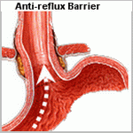
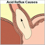
The normal anti-reflux mechanism involves both anatomical and physiological factors: the lower esophageal sphincter, angle of His, crura of the diaphragm, mucosal rosette, swallowed saliva (lubrication and neutralization of acid), antegrade esophageal peristalsis and normal gastric motility/emptying.
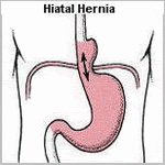
A mild degree of reflux is experienced in most normal individuals, but a major cause of significant GERD is inappropriate transient lower esophageal sphincter relaxations (not preceded by a primary propagated esophageal contraction initiated by swallowing). Other causes include hiatus hernia and delayed gastric emptying.
Although a greater proportion of patients with symptoms of GERD smoke and consume alcohol, clinical studies confirm that traditional risk factors of increasing age, male sex, smoking and alcohol consumption are not strongly associated with symptoms of GERD.
Scope of disease
The majority have mild disease without esophagitis (non-erosive reflux disease). Symptoms can arise with minimal gastric reflux due to increased sensitivity of the esophagus to acid. Moderate disease is associated with esophagitis, and severe chronic disease results in the development of Barrett's esophagus (defined as metaplastic columnar degeneration above the gastro-esophageal junction). This is a premalignant condition, with a 0.5% per patient-year risk of malignant change to adenocarcinoma.
Severe or prolonged reflux can lead to ulceration, bleeding or perforation. Healing occurs by fibrosis and may lead to stricture formation. Severe reflux may also produce a hoarse voice from laryngitis. If the refluxate is aspirated, pneumonia may develop.
Clinical features
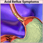
Most patients complain of heartburn, characterized by retrosternal burning pain, precipitated by meals. It may be aggravated by posture (lying flat, stooping, bending forwards) and conditions that raise intra-abdominal pressure (sneezing and coughing). It is often relieved by antacids and may be associated with water brash, the excessive secretion of saliva preceding reflux.
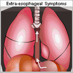
Atypical symptoms include back pain, cardiac-type chest pain, chronic wheeze, nocturnal 'asthma' or recurrent chest infections (due to aspiration), sore throat, hoarse voice, halitosis or dental decay. Dysphagia may occur with esophageal strictures from chronic disease.
Initial investigations
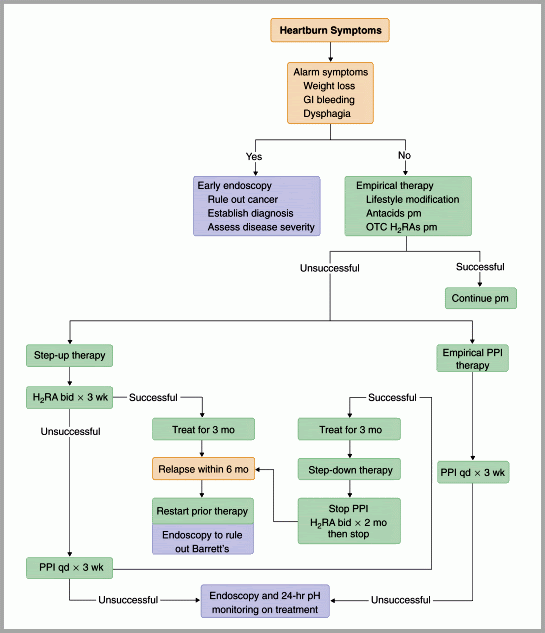
In general, the history can lead to a confident diagnosis of GERD and initial treatment can commence for uncomplicated cases. Older patients, or those who experience weight loss, dysphagia or hematemesis will require further investigations.
Further investigations
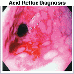
Upper gastrointestinal endoscopy
esophagoscopy is appropriate for patients with symptoms suggestive of complications (weight loss, dysphagia, hematemesis), when the diagnosis is unclear, when symptoms cannot be adequately controlled on medical therapy, or for screening for Barrett's epithelium (in patients over50 with chronic symptoms).
esophageal abnormalities that can be diagnosed at endoscopy include hiatus hernia, esophagitis, Barrett's esophagus, strictures and tumours. The severity of reflux esophagitis can also be assessed at endoscopy, and biopsies can be taken. GERD cannot be excluded by a normal endoscopy.
24-hour pH measurement
Intra-esophageal pH measurement may be required to diagnose GERD if the initial endoscopy is normal or if symptoms do not respond to medical therapy. It is usually performed as an ambulatory test over a 24-hour period, facilitating the diagnosis of GERD by assessment of the duration, frequency and severity of reflux attacks in relation to the pH of the esophagus. It does not provide any information as to the etiology of GERD and may be normal (false negative) in patients with significant bile or volume reflux.
esophageal manometry
Esophageal manometry is a useful investigation to assess esophageal function when planning surgical intervention, especially if achalasia and other esophageal motility disorders are suspected. It is possible to determine the length and position of the lower esophageal sphincter and to measure the sphincter pressure. A short or weak lower esophageal sphincter is often a major contributing cause of reflux esophagitis.
Initial management
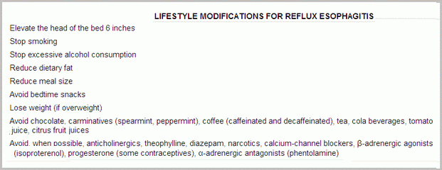
Lifestyle modification
Suggestions that may improve symptoms include weight loss for obese patients, stopping smoking, frequent small meals, avoiding foods that are known to precipitate symptoms, avoiding eating for several hours before bedtime, and raising the head of the bed. However, evidence for improvement associated with these measuresis scarce.
Medical management
There are three pharmacological approaches to controlling symptoms of GERD: neutralization of acid (antacids and alginates), reduction in acid production (H2-receptor blockers, proton pump inhibitors) and increasing gastrointestinal motility (metoclopramide).
Step-up therapy
Most agents for initial therapy are available without prescription, and pharmacists offer the first-line management of GERD in a step-up regimen (starting from the simplest, most cost-effective agent).
Antacids and alginates
Antacids, such as bicarbonate, act by reducing the acidity within the stomach and lower esophagus. Alginate preparations (such as Gaviscon) act by coating the top of the gastric contents to reduce the effect of acid on the esophageal mucosa. Both these agents are effective, but the effects are often short-lived and suitable only for patients with mild symptoms.
H2-receptor antagonists
H2-receptor antagonists (e.g. cimetidine, ranitidine) block the H2-receptors in gastric mucosa, leading to a marked reduction in gastric acid production. Initial symptomatic relief is experienced in approximately 60%.
Step-down therapy
Patients who seek medical attention may already be on a combination of antacids, alginates and H2-receptor antagonists. Current consensus favours step-down therapy (starting with the most effective agent), as it is associated with greater symptomatic relief and fewer treatment failures and physician consultations.
Proton pump inhibitors
The proton pump inhibitors (e.g. omeprazole, lansoprazole) act by blocking the proton pump within the gastric mucosa, almost completely abolishing gastric acid production. Initial symptomatic relief is experienced in approximately 83%.6 An initial 2-4-week course is recommended followed by maintenance therapy. Failure to respond to initial therapy is an indication for further investigations (endoscopy, pH studies) to confirm and assess the severity of the disease.
Prokinetic agents
Due to the superiority of proton pump inhibitors, the role of prokinetic agents (metoclopramide) is ill defined. Prokinetic agents are usually reserved for use in combination with proton pump inhibitor for step-up therapy, or in combination with an H2-receptor blocker for long-term maintenance step-down therapy.
Maintenance therapy
After 6 months of initial therapy 75% of patients experience symptom relapse. A trial of withdrawal of drug therapy may be initiated but the majority will require maintenance therapy either by step-down treatment (long-term H2-receptor blocker) or intermittent on-demand proton pump inhibitor therapy, a 2-4-week course each time symptoms recur.
Surgical management
Before considering anti-reflux surgery, upper gastrointestinal endoscopy, 24-hour pH studies and esophageal manometry are required for confirmatory diagnosis and documentation of the presence and severity of reflux.
Anti-reflux surgery provides effective long-term treatment of GERD with symptomatic control equivalent to medical therapy but lower rates of esophagitis.10 The indications for surgery are failure of medical treatmentor development of complications (ulceration, strictures, Barrett's esophagus or respiratory complications). Relative indications include volume reflux (i.e. excessive volume rather than acid content) and patient preference.
Anti-reflux surgery has a 90% initial success rate. Complications of surgery include dysphagia (usually short-lived)in about 10%, inability to belch, the so-called 'gas bloat syndrome' (20%) and excessive flatus.
Nissen fundoplication
The most common operation is the Nissen (360 degree) fundoplication, which can be performed via an abdominal incision (midline laparotomy) or laparoscopically. The hiatus of the diaphragm is repaired, the lower esophagus is mobilized, then the greater curvature of the stomach via division of the short gastric arteries. The fundus of the stomach is wrapped completely around the lower esophagus.
To reduce the risk of dysphagia, various partial fundoplications have been described, including the anterior partial (Watson), posterior partial (Toupet) and Lind (270 degree) subtotal fundoplications.
Collis-Nissen procedure
This uncommon procedure is reserved for situations where the gastro-esophageal junction cannot be reduced below the diaphragm. An abdominal approach is used and a linear stapler is used to 'lengthen' the esophagus by incorporating the cardia of the stomach prior to creating the wrap.
Management of benign strictures
A barium swallow is often performed to delineate the site and extent of the stricture, followed by endoscopy and biopsies to determine the etiology. The treatment of benign strictures consists of bougie dilatation, and several sessions may be required for resistant strictures. Further treatment options include resection or bypass of the stricture and long-term intubation of the esophagus.
Following esophageal dilatation, the patient is keptnil by mouth if symptoms such as chest or back pain or surgical emphysema develop. A chest film is performed to exclude a pneumomediastinum, which will suggest esophageal perforation.
Prognosis
GERD is a chronic relapsing condition. Long-term studies report 10-year recurrence rates (based on use of anti-reflux medications) in 62% of surgically treated and 92% of medically treated patients. If Barrett's esophagus develops, regular endoscopic surveillance for dysplasia is required.

Monday, March 10, 2008

Acid Reflux Relief logoGreat Videos about Acid Reflux and its Relief

Acid Reflux Video

Saturday, March 8, 2008

Acid Reflux Relief logoHow common is Acid Reflux?

Population-based studies have reported that one third of Western populations experience the symptoms of GERD at least once a month, with 4% to 7% of the population experiencing daily symptoms. Judging from the high prevalence of heartburn in the general population, GERD is a very common condition. Most patients with mild symptoms carry out self-medication, whereas those with more severe and persistent symptoms seek out medical attention. Further, the prevalence and severity of GERD is likely increasing. This is in contrast to duodenal ulcer disease where the prevalence has markedly decreased. These trends may be in part related to the effects of Helicobacter pylori. The diagnosis of a columnar-lined esophagus is also increasing at a rapid rate, and deaths from end-stage benign esophageal disease are on an upward trend. These epidemiologic changes have occurred despite dramatic improvements in the efficacy of treatment options.
Studies on the natural history of GERD are rare. The few that do exist usually involve patients who were receiving some form of therapy. One of the most detailed studies on the natural history of the disease comes from Lausanne, Switzerland, where an intensive endoscopic follow-up of a defined population of 959 patients was performed over a 30-year period. The study involved only patients who had endoscopic esophagitis and did not include those who had symptoms without mucosal injury. It showed that in about 45% of patients esophagitis developed as an isolated episode and does not return while on acid suppression therapy. In the remaining patients esophagitis intermittently recurs on acid suppression therapy, and in 42% it progressed on therapy to more severe mucosal injury. This latter group makes up about 23% of the initial population of patients with esophagitis. The study also showed that 18% of the initial population acquired, while on therapy and within as short a period of 6 weeks, a columnar-lined lower esophagus with intestinal metaplasia.

Friday, March 7, 2008

Acid Reflux Relief logoMedications for Acid Reflux Relief

Medical Treatment of Gastroesophageal Reflux Disease
GERD is such a common condition that most sufferers with mild symptoms carry out self-medication. Sufferers when first seen with symptoms of heartburn without obvious complications can reasonably be placed on 8 to 12 weeks of simple antacids before extensive investigations are carried out. In many situations, this successfully aborts the attacks. Sufferers should be advised to elevate the head of the bed; avoid tight clothing; eat small, frequent meals; avoid eating their nighttime meal shortly before retiring; lose weight; and avoid alcohol, coffee, chocolate, and peppermints, which may aggravate the symptoms. Alginic acid, used in combination with simple antacids, may augment symptomatic relief by creating a physical barrier to reflux as well as by acid reduction. Alginic acid reacts with sodium bicarbonate in the presence of saliva to form a highly viscous solution that floats like a raft on the surface of the gastric contents. When reflux occurs, this protective layer is refluxed into the esophagus and acts as a protective barrier against the noxious gastric contents. Medications to promote gastric emptying, such as metoclopramide, domperidone, or cisapride, are beneficial in early disease but of little value in more severe disease. The mainstay of medical therapy is acid suppression. Sufferers with persistent symptoms should be given hydrogen potassium PPIs, such as omeprazole. In doses as high as 40 mg per day, they can effect an 80% to 90% reduction in gastric acidity. This usually heals mild esophagitis, but healing may occur in only three fourths of sufferers with severe esophagitis. It is important to realize that in sufferers who reflux a combination of gastric and duodenal juice, inadequate acid suppression therapy may give symptomatic improvement while still allowing mixed reflux to occur. This can result in an environment that allows persistent mucosal damage in an asymptomatic sufferer. Unfortunately, within 6 months of discontinuation of any form of medical therapy for GERD, 80% of sufferers have a recurrence of symptoms. In sufferers with reflux disease, esophageal acid exposure is reduced by up to 80% with H2-receptor antagonists and up to 95% with PPIs. Despite the superiority of the latter class of drug over the former, emerging evidence suggests that periods of acid breakthrough still occur. This occurs most commonly at nighttime and is some justification for a split rather than a single dosing regimen. Sufferers with breakthrough reflux symptoms were studied while on omeprazole 20 mg b.i.d. and found that many of them were still refluxing. Intragastric pH monitoring in 28 healthy volunteers and 17 sufferers with reflux disease revealed that nocturnal recovery of acid secretion (more than1 hour) occurred in 75% of the individuals. Recovery of acid secretion occurred within 12 hours of the oral evening dose of PPI, the median recovery time being 7.5 hours. This is particularly pertinent because it is during the nighttime and early morning that asthma symptoms are most pronounced and that peak expiratory flow rate is at its lowest. There have been also shown that ranitidine 300 mg at bedtime is superior to omeprazole 20 mg at bedtime in preventing acid breakthrough. it was speculated to be due to the abolition of histamine-mediated acid secretion in the fasting state. Sufferers presenting for the first time with symptoms suggestive of GE reflux may be given initial therapy with H2 blockers. In view of the availability of these as over-the-counter medication, many sufferers will have already self-medicated their symptoms. Failure of H2 blockers to control the symptoms or immediate return of symptoms after stopping treatment suggests that either the diagnosis is incorrect or the sufferers had relatively severe disease. Endoscopic examination at this stage of the sufferer's evaluation provides the opportunity for assessing the severity of mucosal damage and the presence of Barrett's esophagus. Both of these findings on initial endoscopy predict a high risk for medical failure. A measurement of the degree and pattern of esophageal exposure to gastric and duodenal juice, with 24-hour pH and bilirubin monitoring, should be obtained at this point. The status of the LES and the function of the esophageal body should also be measured. These studies identify features that predict a poor response to medical therapy, frequent relapses, and the development of complications and include supine reflux, poor esophageal contractility, erosive esophagitis or a columnar-lined esophagus at initial presentation, bile in the refluxate, and a structurally defective sphincter. Sufferers who have these risk factors should be given the option of surgery as a primary therapy with the expectation of long-term control of symptoms and complications.

Acid Reflux Relief logoAssessment of esophageal body and gastric function

The presence of poor esophageal body function can impact the likelihood of relief of regurgitation, dysphagia, and respiratory symptoms following surgery and may influence the decision to undertake a partial rather than a complete fundoplication. When peristalsis is absent or severely disordered, many would opt for a partial fundoplication, although recent studies would suggest a complete fundoplication may be appropriate even in this setting. The less favorable response of atypical, compared with typical, reflux symptoms after fundoplication may be related to persistent poor esophageal propulsive function and the continued regurgitation of esophageal contents.
The function of the esophageal body is assessed with esophageal manometry. This is performed with five pressure transducers located in the esophagus. To standardize the procedure the most proximal pressure transducer is located 1 cm below the well-defined cricopharyngeal sphincter. With this method a pressure response along the entire esophagus can be obtained during one swallow. The study consists of recording ten standard wet swallows with 5 mL of water. Amplitude, duration, and morphology of contractions following each swallow are all calculated at the five discrete levels within the esophageal body. The delay between onset or peak of esophageal contractions at the various levels of the esophagus is used to calculate the speed of wave propagation and represents the degree of peristaltic activity.
Esophageal disorders are frequently associated with abnormalities of duodenogastric function. Symptoms suggestive of gastroduodenal pathology include nausea, epigastric pain, anorexia, and early satiety. Abnormalities of gastric motility or increased gastric acid secretion can be responsible for increased esophageal exposure to gastric juice. If not identified before surgery, unrecognized gastric motility abnormalities are occasionally unmasked by an antireflux procedure, resulting in disabling postoperative symptoms. Considerable experience and judgment are necessary to identify the patient with occult gastroduodenal dysfunction. The surgeon should maintain a keen awareness of this possibility and investigate the stomach given any suggestion of problems. Tests of duodenogastric function that are helpful when investigating the patient with GE reflux include gastric emptying studies, gastric acid analysis, 24-hour gastric pH monitoring, and ambulatory bilirubin monitoring of the esophagus and stomach.
Poor gastric emptying or transit can provide for reflux of gastric contents into the distal esophagus. Standard gastric emptying studies are performed with radionuclide-labeled meals. They are often poorly standardized and difficult to interpret. Emptying of solids and liquids can be assessed simultaneously when both phases are marked with different tracers. After ingestion of a labeled standard meal, gamma camera images of the stomach are obtained at 5- to 15-minute intervals for 1.5 to 2 hours.
After correction for decay, the counts in the gastric area are plotted as percentage of total counts at the start of the imaging. The resulting emptying curve can be compared with data obtained in normal volunteers. In general, normal subjects will empty 59% of a meal within 90 minutes.

Acid Reflux Relief logoRadiographic evaluation

Radiographic assessment of the anatomy and function of the esophagus and stomach is one of the most important parts of the preoperative evaluation. Critical issues are assessed, including the presence of esophageal shortening, the size and reducibility of a hiatal hernia, and the propulsive function of the esophagus for both liquids and solids.
The definition of radiographic GE reflux varies depending on whether reflux is spontaneous or induced by various maneuvers. In only about 40% of patients with classic symptoms of GERD is spontaneous reflux observed by the radiologist (i.e., reflux of barium from the stomach into the esophagus with the patient in the upright position). In most patients who show spontaneous reflux on radiography, the diagnosis of increased esophageal acid exposure is confirmed by 24-hour esophageal pH monitoring. Therefore, the radiographic demonstration of spontaneous regurgitation of barium into the esophagus in the upright position is a reliable indicator that reflux is present. Failure to see this does not indicate the absence of disease.
A carefully performed video esophagram can provide an enormous amount of information on the structure and function of the esophagus and stomach. The modern barium swallow emphasizes motion-recording (video), utilizes a tightly controlled examination protocol, and requires an understanding of esophageal physiology.
Videotaping the study greatly aids the evaluation, providing the surgeon with a real-time assessment of swallowing function, bolus transport, and the size and reducibility of hiatal hernias. Given routine review before antireflux surgery, its value becomes increasingly clear. The study provides structural information including the presence of obstructing lesions and anatomic abnormalities of the foregut. A hiatal hernia is present in more than 80% of patients with GE reflux and is best demonstrated with the patient in the prone position, which causes increased abdominal pressure and promotes distention of the hernia above the diaphragm. The presence of a hiatal hernia is an important component of the underlying pathophysiology of GE reflux. Other relevant findings include a large (greater than 5 cm) or irreducible hernia, suggesting the presence of a shortened esophagus; a tight crural collar that inhibits barium transit into the stomach, suggesting a possible cause of dysphagia; and the presence of a paraesophageal hernia.
Lower esophageal narrowing resulting from a ring, stricture, or obstructing lesion is optimally viewed with full distention of the esophagogastric region. A full-column technique with distention of the esophageal wall can be used to discern extrinsic compression of the esophagus. Mucosal relief or double-contrast films should be obtained to enhance the detection of small esophageal neoplasms, mild esophagitis, and esophageal varices. The pharynx and upper esophageal sphincter are evaluated in the upright position, and an assessment of the relative timing and coordination of pharyngeal transit is possible.
The assessment of peristalsis on video esophagram often adds to, or complements, the information obtained by esophageal motility studies. This is in part because the video barium study can be done both upright and supine and with liquid and solid bolus material, which is not true of a stationary motility examination. This is particularly true with subtle motility abnormalities. During normal swallowing, a stripping wave (primary peristalsis) is generated that completely clears the bolus. Residual material can stimulate a secondary peristaltic wave, but usually a second pharyngeal swallow is required. Motility disorders with disorganized or simultaneous esophageal contractions have tertiary waves and provide a segmented appearance to the barium column, often referred to as beading or corkscrewing. In dysphagic patients, a barium-impregnated marshmallow, bread, or hamburger is a useful adjunct, which can discern a functional esophageal transport disturbance not evident on the liquid barium study. Reflux is not easily seen on video esophagram, and motility disorders that cause retrograde barium transport may be mistaken for reflux.
Assessment of the stomach and duodenum during the barium study is a necessity for proper preoperative evaluation of the patient with GERD. Evidence of gastric or duodenal ulcer, neoplasm, or poor gastroduodenal transit has obvious importance in the proper preoperative evaluation.

Acid Reflux Relief logoAssessment of esophageal length

Esophageal shortening is a consequence of scarring and fibrosis associated with repetitive esophageal injury. Anatomic shortening of the esophagus can compromise the ability to perform an adequate tension-free fundoplication and may result in an increased incidence of breakdown or thoracic displacement of the repair. Esophageal length is best assessed preoperatively using video roentgenographic contrast studies and endoscopic findings. Endoscopically, hernia size is measured as the difference between the diaphragmatic crura, identified by having the patient sniff, and the GE junction, identified as the loss of gastric rugal folds. We consider the possibility of a short esophagus in patients with strictures or those with large hiatal hernias (greater than 5 cm), particularly when the latter fail to reduce in the upright position on a video barium esophagram.
The definitive determination of esophageal shortening is made intraoperatively when, after thorough mobilization of the esophagus, the GE junction cannot be reduced below the diaphragmatic hiatus without undue tension on the esophageal body. Surgeons performing fundoplication have reported varying incidences of esophageal shortening, attesting to the judgment inherent in defining and recognizing undue tension. An advantage of transthoracic fundoplication is the ability to mobilize the esophagus extensively from the diaphragmatic hiatus to the aortic arch. With the GE junction marked with a suture, esophageal shortening is defined by an inability to position the repair beneath the diaphragm without tension. In this situation, a Collis gastroplasty coupled with either a partial or complete fundoplication may be performed.
Potential pitfalls of laparoscopic fundoplication include the elevation of the diaphragm due to pneumoperitoneum, potentially contributing to a false impression that esophageal length is adequate, and the limited ability to mobilize the esophagus relative to the transthoracic approach. In our experience, the failure to appreciate esophageal shortening is a major cause of fundoplication failure and is often the explanation for the slipped Nissen fundoplication. In many such instances, the initial repair is incorrectly constructed around the proximal tubularized stomach rather than the terminal esophagus. Surgeons opting to perform fundoplication laparoscopically in the setting of potential esophageal shortening must be vigilant of esophageal tension, technically facile at extensive mediastinal mobilization of the esophagus while preserving vagal integrity, and able to perform a laparoscopic or open transabdominal Collis gastroplasty should esophageal lengthening be necessary.

Acid Reflux Relief logoTwenty-four hour ambulatory pH monitoring

The most direct method of assessing the relationship between symptoms and GERD is to measure the esophageal exposure to gastric juice with an indwelling pH electrode. Miller first reported prolonged esophageal pH monitoring in 1964, although it was not until 1973 that its clinical applicability and advantages were demonstrated by Johnson and DeMeester. Ambulatory pH testing is considered by many to be the gold standard for the diagnosis of GERD, because it has the highest sensitivity and specificity of all tests currently available. Some experts have suggested that 24-hour pH monitoring be used selectively, limited to patients with atypical symptoms or no endoscopic evidence of GE reflux. Given present-day referral patterns, more than half of the patients referred for antireflux surgery will have no endoscopic evidence of mucosal injury. For these patients, 24-hour pH monitoring provides the only objective measure of the presence of pathologic esophageal acid exposure. Although it is true that most patients with typical symptoms and erosive esophagitis have a positive 24-hour pH result, the study provides other useful information. It quantifies the actual time that the esophageal mucosa is exposed to gastric juice, measures the ability of the esophagus to clear refluxed acid and correlates esophageal acid exposure with the patient's symptoms. It is the only way to quantitatively express the overall degree and pattern of esophageal acid exposure, both of which may impact the decision toward surgery. Patients with nocturnal or bipositional reflux have a higher prevalence of complications and failure of long-term medical control. For these reasons, we continue to advocate its routine use in clinical practice.
The units used to express esophageal exposure to gastric juice are (a) cumulative time the esophageal pH is below a chosen threshold, expressed as the percent of the total, upright, and supine monitored time; (b) frequency of reflux episodes below a chosen threshold, expressed as number of episodes per 24 hours; and (c) duration of the episodes, expressed as the number of episodes greater than 5 minutes per 24 hours and the time in minutes of the longest episode recorded. The upper limits of normal were established at the 95th percentile. Most centers use pH 4 as the threshold. Combining the result of the six components into one expression that reflects the overall esophageal acid exposure below a pH threshold, a pH score was calculated by using the standard deviation of the mean of each of the six components measured.

Acid Reflux Relief logoEndoscopic evaluation

Endoscopic visualization of the esophagus equates to the physical examination of the foregut and is a critical part of the preoperative evaluation of patients with GERD. Its main aim is to detect complications of GE reflux, the presence of which may influence therapeutic decisions.
In every patient, the locations of the diaphragmatic crura, the GE junction, and the squamocolumnar junction are determined. These anatomic landmarks are commonly at three different sites in patients with GERD. The crura are usually evident and can be confirmed by having the patient sniff during the examination. The anatomic GE junction is identified as the point where the gastric rugal folds meet the tubular esophagus and is often below the squamocolumnar junction, even in patients without otherwise obvious Barrett's esophagus.
Endoscopic esophagitis is defined by the presence of mucosal erosions . When present, the grade and length of esophageal mucosal injury are recorded. The presence and length of columnar epithelium extending above the anatomic GE junction is also noted. It is suspected at endoscopy when there is difficulty in visualizing the squamocolumnar junction at its normal location and by the appearance of a velvety red luxuriant mucosa. The presence of Barrett's esophagus is confirmed by biopsy evidence of specialized intestinal metaplasia and is considered histologic evidence of GERD. Endoscopic visualization of columnar lining without histologic confirmation of specialized intestinal metaplasia is not considered Barrett's esophagus and likely has no premalignant potential. Multiple biopsies should be taken in a cephalad direction to determine the level at which the junction of Barrett's epithelium and normal squamous mucosa occurs. Barrett's esophagus is susceptible to ulceration, bleeding, stricture formation, and malignant degeneration. Dysplasia is the earliest sign of malignant change. Because dysplastic changes typically occur in a random distribution within the distal esophagus, a minimum of four biopsies (each quadrant) every 2 cm should be obtained from the metaplastic epithelium. Particular attention must be paid to the squamocolumnar junction in these patients, where a mass, ulcer, nodularity, or inflammatory tissue is always considered suspicious for malignancy and requires thorough biopsy. The GE junction is defined endoscopically where the tubular esophagus meets gastric rugal folds, and the squamocolumnar junction is where there is an obvious change from the velvety and darker columnar epithelium to the lighter squamous epithelium.
After completion of the esophageal examination, the first and second portions of the duodenum and the stomach are systematically inspected. This is commonly done on withdrawal of the endoscope. When the antrum is visualized, the incisura angularis appears as a constant ridge on the lesser curve. Turning the lens of the scope 180 degrees allows inspection of the fundus and cardia. Attention is paid to the frenulum (angle of His) of the esophagogastric junction and to the closeness with which the cardia grips the scope. The appearance of this valve have been graded on a scale from I to IV according to the degree of unfolding or deterioration of the normal valve architecture. This grading system has been correlated with the presence of increased esophageal acid exposure, occurring predominantly in patients with a grade III or IV valve.
A hiatal hernia is endoscopically confirmed by finding a pouch lined with gastric rugal folds lying 2 cm or more above the margins of the diaphragmatic crura. A prominent sliding hernia is frequently associated with increased esophageal exposure to gastric juice. When a paraesophageal hernia exists, particular attention is given to exclude a gastric ulcer or gastritis within the pouch. The intragastric retroflex or J maneuver is important in evaluating the full circumference of the mucosal lining of the herniated stomach. As the endoscope is removed, the esophagus is again examined and biopsies taken. The location of the cricopharyngeus is identified and the larynx and vocal cords are visualized. Acid reflux may result in inflammation of the larynx. Vocal cord movement is recorded both as a reference for subsequent surgery and an assessment of the patient's ability to protect the airway.

Acid Reflux Relief logoSurgery for Acid Reflux Relief

Indications of Antireflux Surgery
Antireflux surgery is indicated for the treatment of objectively documented, relatively severe GERD. Candidates for surgery include not only patients with erosive esophagitis, stricture, and Barrett's esophagus but also those without severe mucosal injury who are dependent on PPIs for symptom relief. Patients with atypical or respiratory symptoms who have a good response to intensive medical treatment are also candidates. The option of antireflux surgery should be given to all patients who have demonstrated the need for long-term medical therapy, particularly if escalating doses of PPIs are needed to control symptoms. Antireflux surgery may be the preferred option in patients younger than 50 years, those who are noncompliant with their drug regimen, those for whom medications are a financial burden, and those who favor a single intervention over long-term drug treatment. It may be the treatment of choice in patients who are at high risk of progression despite medical therapy. Although this population is not well defined, risk factors that predict progressive disease and a poor response to medical therapy include (a) nocturnal reflux on 24-hour esophageal pH study, (b) a structurally deficient LES, (c) mixed reflux of gastric and duodenal juice, and (d) mucosal injury at presentation.

Preoperative Evaluation
Successful antireflux surgery is largely defined by two objectives: the achievement of long-term relief of reflux symptoms and the absence of complications or complaints after the operation. In practice, achieving these two deceptively simple goals is difficult. Both are critically dependent on establishing that the symptoms for which the operation is performed are the result of excess esophageal exposure to gastric juice, as well as the proper performance of the appropriate antireflux procedure. Success can be expected in the vast majority of patients if these two criteria are met. The status of the LES is not as important a factor as in the days of open surgery. Patients with normal resting sphincters are often selected for antireflux surgery in the era of laparoscopic fundoplication. The outcome is not dependent on sphincter function.
There are four important goals of the diagnostic approach to patients suspected of having GERD and being considered for antireflux surgery.

Objective Documentation
The introduction of laparoscopic access, coupled with the growing recognition that surgery is a safe and durable treatment for GERD, has dramatically increased the number of patients being referred for laparoscopic fundoplication. The threshold for surgical referral is such that increasing numbers of patients without endoscopic esophagitis or other objective evidence of the presence of reflux are now considered candidates for laparoscopic antireflux surgery. These facts combine to underscore the importance of selecting patients for surgery who are likely to have a successful outcome. Although a Nissen fundoplication will reliably and reproducibly halt the return of gastroduodenal juice into the esophagus, little benefit is likely if the patient's symptoms are not caused by this specific pathophysiologic derangement. Thus, in large part, the anticipated success rate of laparoscopic fundoplication is directly proportional to the degree of certainty that GERD is the underlying cause of the patient's complaints.
Three factors predictive of a successful outcome following antireflux surgery have emerged . These are (a) an abnormal score on 24-hour esophageal pH monitoring; (b) the presence of typical symptoms of GERD, namely heartburn or regurgitation; and (c) symptomatic improvement in response to acid suppression therapy prior to surgery. It is immediately evident that each of these factors helps to establish that GERD is indeed the cause of the patient's symptoms and that they have little to do with the severity of the disease.

Endoscopic evaluation

Twenty-four hour ambulatory pH monitoring

Assessment of esophageal length

Radiographic evaluation

Assessment of esophageal body and gastric function